 Movie
Movie Controller


+ Open data

- Basic information
| Entry | Database: PDB / ID: 5t46 | ||||||
|---|---|---|---|---|---|---|---|
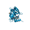
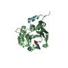
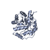
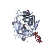
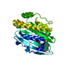
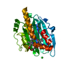
| Title | Crystal structure of the human eIF4E-eIF4G complex | ||||||
 Components Components |
| ||||||
 Keywords Keywords | TRANSLATION / GENE REGULATION / CAP BINDING PROTEIN / 4E-BINDING PROTEIN / TRANSLATION INITIATION / eIF4F | ||||||
| Function / homology |  Function and homology information Function and homology informationpositive regulation of eukaryotic translation initiation factor 4F complex assembly / positive regulation of translation in response to endoplasmic reticulum stress / eukaryotic initiation factor 4G binding / Activation of the mRNA upon binding of the cap-binding complex and eIFs, and subsequent binding to 43S / eukaryotic initiation factor 4E binding / cap-dependent translational initiation / regulation of translation at postsynapse, modulating synaptic transmission / RNA cap binding / eukaryotic translation initiation factor 4F complex / Z-decay: degradation of maternal mRNAs by zygotically expressed factors ...positive regulation of eukaryotic translation initiation factor 4F complex assembly / positive regulation of translation in response to endoplasmic reticulum stress / eukaryotic initiation factor 4G binding / Activation of the mRNA upon binding of the cap-binding complex and eIFs, and subsequent binding to 43S / eukaryotic initiation factor 4E binding / cap-dependent translational initiation / regulation of translation at postsynapse, modulating synaptic transmission / RNA cap binding / eukaryotic translation initiation factor 4F complex / Z-decay: degradation of maternal mRNAs by zygotically expressed factors / chromatoid body / regulation of cellular response to stress / translation factor activity, RNA binding / Deadenylation of mRNA / mRNA cap binding / Transport of the SLBP independent Mature mRNA / Transport of the SLBP Dependant Mature mRNA / miRNA-mediated gene silencing by inhibition of translation / RNA 7-methylguanosine cap binding / Transport of Mature mRNA Derived from an Intronless Transcript / M-decay: degradation of maternal mRNAs by maternally stored factors / RISC complex / regulation of translational initiation / positive regulation of protein localization to cell periphery / negative regulation of neuron differentiation / stem cell population maintenance / Ribosomal scanning and start codon recognition / Translation initiation complex formation / positive regulation of protein metabolic process / mTORC1-mediated signalling / cellular response to dexamethasone stimulus / regulation of presynapse assembly / Nonsense Mediated Decay (NMD) independent of the Exon Junction Complex (EJC) / GTP hydrolysis and joining of the 60S ribosomal subunit / positive regulation of G1/S transition of mitotic cell cycle / L13a-mediated translational silencing of Ceruloplasmin expression / mRNA export from nucleus / behavioral fear response / Nonsense Mediated Decay (NMD) enhanced by the Exon Junction Complex (EJC) / energy homeostasis / translation initiation factor activity / positive regulation of neuron differentiation / translation initiation factor binding / positive regulation of mitotic cell cycle / cellular response to nutrient levels / negative regulation of autophagy / P-body / AUF1 (hnRNP D0) binds and destabilizes mRNA / translational initiation / G1/S transition of mitotic cell cycle / ISG15 antiviral mechanism / neuron differentiation / Regulation of expression of SLITs and ROBOs / cytoplasmic stress granule / cytoplasmic ribonucleoprotein granule / regulation of translation / positive regulation of cell growth / DNA-binding transcription factor binding / molecular adaptor activity / negative regulation of translation / nuclear speck / postsynapse / ribosome / translation / mRNA binding / perinuclear region of cytoplasm / glutamatergic synapse / enzyme binding / RNA binding / extracellular exosome / ATP binding / membrane / nucleus / cytoplasm / cytosol Similarity search - Function | ||||||
| Biological species |  Homo sapiens (human) Homo sapiens (human) | ||||||
| Method |  X-RAY DIFFRACTION / SYNCHROTRON / MOLECULAR REPLACEMENT / Resolution: 1.53 Å X-RAY DIFFRACTION / SYNCHROTRON / MOLECULAR REPLACEMENT / Resolution: 1.53 Å | ||||||
 Authors Authors | Gruener, S. / Peter, D. / Weber, R. / Wohlbold, L. / Chung, M.-Y. / Weichenrieder, O. / Valkov, E. / Igreja, C. / Izaurralde, E. | ||||||
 Citation Citation | Journal: Mol.Cell / Year: 2016 Title: The Structures of eIF4E-eIF4G Complexes Reveal an Extended Interface to Regulate Translation Initiation. Authors: Gruner, S. / Peter, D. / Weber, R. / Wohlbold, L. / Chung, M.Y. / Weichenrieder, O. / Valkov, E. / Igreja, C. / Izaurralde, E. | ||||||
| History |
|
- Structure visualization
| Structure viewer | Molecule: MolmilJmol/JSmol |
|---|
- Downloads & links
-Download
| PDBx/mmCIF format | 5t46.cif.gz | 284 KB | Display | PDBx/mmCIF format |
|---|---|---|---|---|
| PDB format | pdb5t46.ent.gz | 234.5 KB | Display | PDB format |
| PDBx/mmJSON format | 5t46.json.gz | Tree view | PDBx/mmJSON format | |
| Others |  Other downloads Other downloads |
-Validation report
| Arichive directory | https://data.pdbj.org/pub/pdb/validation_reports/t4/5t46ftp://data.pdbj.org/pub/pdb/validation_reports/t4/5t46 | HTTPS FTP |
|---|
-Related structure data
| Related structure data |  5t47C 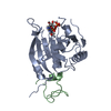 5t48C 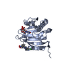 4tpwS S: Starting model for refinement C: citing same article ( |
|---|---|
| Similar structure data |


-Links
 PDBj
PDBj


- Assembly
| Deposited unit | 
| ||||||||
|---|---|---|---|---|---|---|---|---|---|
| 1 | 
| ||||||||
| 2 | 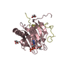
| ||||||||
| Unit cell |
|
-Components
| #1: Protein | Mass: 25503.711 Da / Num. of mol.: 2 Source method: isolated from a genetically manipulated source Source: (gene. exp.) Homo sapiens (human) / Gene: EIF4E, EIF4EL1, EIF4F / Plasmid: PETMCN / Details (production host): (PNYC) / Production host:  #2: Protein | Mass: 7876.048 Da / Num. of mol.: 2 Source method: isolated from a genetically manipulated source Source: (gene. exp.) Homo sapiens (human) / Gene: EIF4G1, EIF4F, EIF4G, EIF4GI / Plasmid: PETMCN / Details (production host): (PNEA) / Production host: #3: Chemical |   Mass: 538.215 Da / Num. of mol.: 2 / Source method: obtained synthetically / Formula: C11H19N5O14P3 Mass: 538.215 Da / Num. of mol.: 2 / Source method: obtained synthetically / Formula: C11H19N5O14P3#4: Chemical | ChemComp-GOL /   Mass: 92.094 Da / Num. of mol.: 7 / Source method: obtained synthetically / Formula: C3H8O3 Mass: 92.094 Da / Num. of mol.: 7 / Source method: obtained synthetically / Formula: C3H8O3#5: Water | ChemComp-HOH / |  Mass: 18.015 Da / Num. of mol.: 346 / Source method: isolated from a natural source / Formula: H2O Mass: 18.015 Da / Num. of mol.: 346 / Source method: isolated from a natural source / Formula: H2O |
|---|
-Experimental details
-Experiment
| Experiment | Method: X-RAY DIFFRACTION / Number of used crystals: 1 |
|---|
- Sample preparation
| Crystal | Density Matthews: 1.9 Å3/Da / Density % sol: 35 % |
|---|---|
| Crystal grow | Temperature: 293 K / Method: vapor diffusion, sitting drop / pH: 7.25 / Details: 0.1 M Hepes pH 7.25, 25% w/v PEG 6000 |
-Data collection
| Diffraction | Mean temperature: 100 K |
|---|---|
| Diffraction source | Source: SYNCHROTRON / Site: SOLEIL  / Beamline: PROXIMA 1 / Wavelength: 0.97857 Å / Beamline: PROXIMA 1 / Wavelength: 0.97857 Å |
| Detector | Type: DECTRIS PILATUS 6M / Detector: PIXEL / Date: Jul 23, 2015 / Details: DYNAMICALLY BENDABLE MIRRORS |
| Radiation | Monochromator: SI(111) / Protocol: SINGLE WAVELENGTH / Monochromatic (M) / Laue (L): M / Scattering type: x-ray |
| Radiation wavelength | Wavelength: 0.97857 Å / Relative weight: 1 |
| Reflection | Resolution: 1.53→44.65 Å / Num. obs: 74453 / % possible obs: 99.8 % / Observed criterion σ(I): -3 / Redundancy: 3.7 % / Biso Wilson estimate: 25 Å2 / CC1/2: 0.998 / Rsym value: 0.052 / Net I/σ(I): 11.6 |
| Reflection shell | Resolution: 1.53→1.56 Å / Redundancy: 3.8 % / Rmerge(I) obs: 1.013 / Mean I/σ(I) obs: 1.2 / CC1/2: 0.565 / Rsym value: 1.013 / % possible all: 99.9 |
- Processing
| Software |
| |||||||||||||||||||||||||||||||||||||||||||||||||||||||||||||||||||||||||||||||||||||||||||||||||||||||||||||||||||||||||||||||||||||||||||||||||||||||||||||||||||||||||||||||||||||||||||||
|---|---|---|---|---|---|---|---|---|---|---|---|---|---|---|---|---|---|---|---|---|---|---|---|---|---|---|---|---|---|---|---|---|---|---|---|---|---|---|---|---|---|---|---|---|---|---|---|---|---|---|---|---|---|---|---|---|---|---|---|---|---|---|---|---|---|---|---|---|---|---|---|---|---|---|---|---|---|---|---|---|---|---|---|---|---|---|---|---|---|---|---|---|---|---|---|---|---|---|---|---|---|---|---|---|---|---|---|---|---|---|---|---|---|---|---|---|---|---|---|---|---|---|---|---|---|---|---|---|---|---|---|---|---|---|---|---|---|---|---|---|---|---|---|---|---|---|---|---|---|---|---|---|---|---|---|---|---|---|---|---|---|---|---|---|---|---|---|---|---|---|---|---|---|---|---|---|---|---|---|---|---|---|---|---|---|---|---|---|---|---|
| Refinement | Method to determine structure: MOLECULAR REPLACEMENT Starting model: 4TPW Resolution: 1.53→44.647 Å / SU ML: 0.17 / Cross valid method: FREE R-VALUE / σ(F): 1.01 / Phase error: 16.69 Details: TORSIONAL NCS RESTRAINTS WERE APPLIED RELATING CHAINS A, B TO CHAINS C, D. INDIVIDUAL B-FACTORS WERE REFINED ANISOTROPICALLY EXCEPT FOR WATER ATOMS. HYDROGENS WERE REFINED IN THE RIDING POSITIONS
| |||||||||||||||||||||||||||||||||||||||||||||||||||||||||||||||||||||||||||||||||||||||||||||||||||||||||||||||||||||||||||||||||||||||||||||||||||||||||||||||||||||||||||||||||||||||||||||
| Solvent computation | Shrinkage radii: 0.9 Å / VDW probe radii: 1.11 Å | |||||||||||||||||||||||||||||||||||||||||||||||||||||||||||||||||||||||||||||||||||||||||||||||||||||||||||||||||||||||||||||||||||||||||||||||||||||||||||||||||||||||||||||||||||||||||||||
| Displacement parameters | Biso mean: 34.99 Å2 | |||||||||||||||||||||||||||||||||||||||||||||||||||||||||||||||||||||||||||||||||||||||||||||||||||||||||||||||||||||||||||||||||||||||||||||||||||||||||||||||||||||||||||||||||||||||||||||
| Refinement step | Cycle: LAST / Resolution: 1.53→44.647 Å
| |||||||||||||||||||||||||||||||||||||||||||||||||||||||||||||||||||||||||||||||||||||||||||||||||||||||||||||||||||||||||||||||||||||||||||||||||||||||||||||||||||||||||||||||||||||||||||||
| Refine LS restraints |
| |||||||||||||||||||||||||||||||||||||||||||||||||||||||||||||||||||||||||||||||||||||||||||||||||||||||||||||||||||||||||||||||||||||||||||||||||||||||||||||||||||||||||||||||||||||||||||||
| LS refinement shell |
|
