 Movie
Movie Controller

[English] 日本語
 Yorodumi
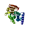
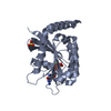
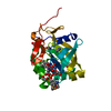
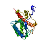
- PDB-1r18: Drosophila protein isoaspartyl methyltransferase with S-adenosyl-... -
+ Open data

- Basic information
| Entry | Database: PDB / ID: 1r18 | ||||||
|---|---|---|---|---|---|---|---|
| Title | Drosophila protein isoaspartyl methyltransferase with S-adenosyl-L-homocysteine | ||||||
 Components Components | Protein-L-isoaspartate(D-aspartate)-O-methyltransferase | ||||||
 Keywords Keywords | TRANSFERASE / methyltransferase / isomerization / protein repair / S-adenosyl homocysteine | ||||||
| Function / homology |  Function and homology information Function and homology informationProtein repair / protein-L-isoaspartate(D-aspartate) O-methyltransferase / protein-L-isoaspartate (D-aspartate) O-methyltransferase activity / methylation / defense response to bacterium / cytoplasm / cytosol Similarity search - Function | ||||||
| Biological species |  | ||||||
| Method |  X-RAY DIFFRACTION / MOLECULAR REPLACEMENT / Resolution: 2.2 Å X-RAY DIFFRACTION / MOLECULAR REPLACEMENT / Resolution: 2.2 Å | ||||||
 Authors Authors | Bennett, E.J. / Bjerregaard, J. / Knapp, J.E. / Chavous, D.A. / Friedman, A.M. / Royer Jr., W.E. / O'Connor, C.M. | ||||||
 Citation Citation | Journal: Biochemistry / Year: 2003 Title: Catalytic implications from the Drosophila protein L-isoaspartyl methyltransferase structure and site-directed mutagenesis. Authors: Bennett, E.J. / Bjerregaard, J. / Knapp, J.E. / Chavous, D.A. / Friedman, A.M. / Royer Jr., W.E. / O'Connor, C.M. #1: Journal: Protein Sci. / Year: 2002Title: Crystal structure of human L-isoaspartyl-O-methyl-transferase with S-adenosyl homocysteine at 1.6A resolution and modeling of an isoaspartyl-containing peptide at the active site Authors: Smith, C.D. / Carson, M. / Friedman, A.M. / Skinner, M.M. / Delucas, L. / Chantalat, L. / Weise, L. / Shirasawa, T. / Chattopadhyay, D. | ||||||
| History |
|
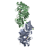
- Structure visualization
| Structure viewer | Molecule: MolmilJmol/JSmol |
|---|
- Downloads & links
-Download
| PDBx/mmCIF format | 1r18.cif.gz | 55.7 KB | Display | PDBx/mmCIF format |
|---|---|---|---|---|
| PDB format | pdb1r18.ent.gz | 39.1 KB | Display | PDB format |
| PDBx/mmJSON format | 1r18.json.gz | Tree view | PDBx/mmJSON format | |
| Others |  Other downloads Other downloads |
-Validation report
| Arichive directory | https://data.pdbj.org/pub/pdb/validation_reports/r1/1r18ftp://data.pdbj.org/pub/pdb/validation_reports/r1/1r18 | HTTPS FTP |
|---|
-Related structure data
| Related structure data | 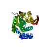 1i1nS S: Starting model for refinement |
|---|---|
| Similar structure data |


-Links
 PDBj
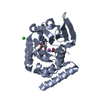
PDBj- Assembly
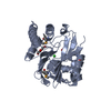
| Deposited unit | 
| ||||||||
|---|---|---|---|---|---|---|---|---|---|
| 1 |
| ||||||||
| 2 | 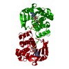
| ||||||||
| Unit cell |
|
-Components
| #1: Protein | Mass: 24597.859 Da / Num. of mol.: 1 Source method: isolated from a genetically manipulated source Source: (gene. exp.)  References: UniProt: Q27869, protein-L-isoaspartate(D-aspartate) O-methyltransferase |
|---|---|
| #2: Chemical | ChemComp-SAH /   Type: L-peptide linking / Mass: 384.411 Da / Num. of mol.: 1 / Source method: obtained synthetically / Formula: C14H20N6O5S Type: L-peptide linking / Mass: 384.411 Da / Num. of mol.: 1 / Source method: obtained synthetically / Formula: C14H20N6O5S |
| #3: Water | ChemComp-HOH /  Mass: 18.015 Da / Num. of mol.: 77 / Source method: isolated from a natural source / Formula: H2O Mass: 18.015 Da / Num. of mol.: 77 / Source method: isolated from a natural source / Formula: H2O |
-Experimental details
-Experiment
| Experiment | Method: X-RAY DIFFRACTION / Number of used crystals: 1 |
|---|
- Sample preparation
| Crystal | Density Matthews: 1.98 Å3/Da / Density % sol: 37.79 % | ||||||||||||||||||||||||
|---|---|---|---|---|---|---|---|---|---|---|---|---|---|---|---|---|---|---|---|---|---|---|---|---|---|
| Crystal grow | Temperature: 277 K / Method: vapor diffusion, sitting drop / pH: 7.5 Details: 100mM Tris, 35% v/v Ethanol, pH 7.5, VAPOR DIFFUSION, SITTING DROP, temperature 277K | ||||||||||||||||||||||||
| Crystal grow | *PLUS Temperature: 4 ℃ / Method: vapor diffusion, sitting drop | ||||||||||||||||||||||||
| Components of the solutions | *PLUS
|
-Data collection
| Diffraction | Mean temperature: 298 K |
|---|---|
| Diffraction source | Source: ROTATING ANODE / Type: RIGAKU RU300 / Wavelength: 1.5418 Å |
| Detector | Type: RIGAKU RAXIS IV / Detector: IMAGE PLATE / Date: Apr 12, 2002 / Details: osmic mirrors |
| Radiation | Monochromator: Osmic mirrors / Protocol: SINGLE WAVELENGTH / Monochromatic (M) / Laue (L): M / Scattering type: x-ray |
| Radiation wavelength | Wavelength: 1.5418 Å / Relative weight: 1 |
| Reflection | Resolution: 2.2→40 Å / Num. all: 8636 / Num. obs: 8636 / % possible obs: 89 % / Observed criterion σ(F): 1 / Observed criterion σ(I): 1 / Redundancy: 1.7 % / Biso Wilson estimate: 10.4 Å2 / Rmerge(I) obs: 0.088 / Rsym value: 0.088 / Net I/σ(I): 11.4 |
| Reflection shell | Resolution: 2.2→2.28 Å / Redundancy: 1.3 % / Rmerge(I) obs: 0.232 / Num. unique all: 761 / Rsym value: 0.232 / % possible all: 79.6 |
| Reflection | *PLUS % possible obs: 89 % / Num. measured all: 14628 |
| Reflection shell | *PLUS Highest resolution: 2.2 Å / % possible obs: 79.6 % / Num. unique obs: 761 / Mean I/σ(I) obs: 2.9 |
- Processing
| Software |
| ||||||||||||||||||||||||||||||||||||||||||||||||||||||||||||
|---|---|---|---|---|---|---|---|---|---|---|---|---|---|---|---|---|---|---|---|---|---|---|---|---|---|---|---|---|---|---|---|---|---|---|---|---|---|---|---|---|---|---|---|---|---|---|---|---|---|---|---|---|---|---|---|---|---|---|---|---|---|
| Refinement | Method to determine structure: MOLECULAR REPLACEMENT Starting model: Human PIMT (pdb code 1I1N) Resolution: 2.2→20.97 Å / Rfactor Rfree error: 0.011 / Isotropic thermal model: GROUP / Cross valid method: THROUGHOUT / σ(F): 1 / Stereochemistry target values: Engh & Huber
| ||||||||||||||||||||||||||||||||||||||||||||||||||||||||||||
| Solvent computation | Solvent model: FLAT MODEL / Bsol: 55.122 Å2 / ksol: 0.318386 e/Å3 | ||||||||||||||||||||||||||||||||||||||||||||||||||||||||||||
| Displacement parameters | Biso mean: 23 Å2
| ||||||||||||||||||||||||||||||||||||||||||||||||||||||||||||
| Refine analyze |
| ||||||||||||||||||||||||||||||||||||||||||||||||||||||||||||
| Refinement step | Cycle: LAST / Resolution: 2.2→20.97 Å
| ||||||||||||||||||||||||||||||||||||||||||||||||||||||||||||
| Refine LS restraints |
| ||||||||||||||||||||||||||||||||||||||||||||||||||||||||||||
| LS refinement shell | Resolution: 2.2→2.28 Å / Rfactor Rfree error: 0.043 / Total num. of bins used: 10
| ||||||||||||||||||||||||||||||||||||||||||||||||||||||||||||
| Xplor file |
| ||||||||||||||||||||||||||||||||||||||||||||||||||||||||||||
| Refinement | *PLUS Highest resolution: 2.2 Å / Lowest resolution: 40 Å | ||||||||||||||||||||||||||||||||||||||||||||||||||||||||||||
| Solvent computation | *PLUS | ||||||||||||||||||||||||||||||||||||||||||||||||||||||||||||
| Displacement parameters | *PLUS | ||||||||||||||||||||||||||||||||||||||||||||||||||||||||||||
| Refine LS restraints | *PLUS
|
