 Movie
Movie Controller

[English] 日本語
 Yorodumi
- PDB-1i1n: HUMAN PROTEIN L-ISOASPARTATE O-METHYLTRANSFERASE WITH S-ADENOSYL ... -
+ Open data

- Basic information
| Entry | Database: PDB / ID: 1i1n | ||||||
|---|---|---|---|---|---|---|---|
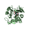
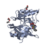
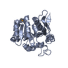
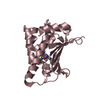
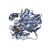
| Title | HUMAN PROTEIN L-ISOASPARTATE O-METHYLTRANSFERASE WITH S-ADENOSYL HOMOCYSTEINE | ||||||
 Components Components | PROTEIN-L-ISOASPARTATE O-METHYLTRANSFERASE | ||||||
 Keywords Keywords | TRANSFERASE / METHYLTRANSFERASE / S-ADENOSYL HOMOCYSTEINE / PROTEIN REPAIR | ||||||
| Function / homology |  Function and homology information Function and homology informationprotein-L-isoaspartate(D-aspartate) O-methyltransferase / protein-L-isoaspartate (D-aspartate) O-methyltransferase activity / Protein repair / protein repair / protein methylation / extracellular vesicle / cadherin binding / extracellular exosome / cytoplasm / cytosol Similarity search - Function | ||||||
| Biological species |  Homo sapiens (human) Homo sapiens (human) | ||||||
| Method |  X-RAY DIFFRACTION / MOLECULAR REPLACEMENT / Resolution: 1.5 Å X-RAY DIFFRACTION / MOLECULAR REPLACEMENT / Resolution: 1.5 Å | ||||||
 Authors Authors | Smith, C.D. / Chattopadhyay, D. / Carson, M. / Friedman, A.M. / Skinner, M.M. | ||||||
 Citation Citation | Journal: Protein Sci. / Year: 2002 Title: Crystal structure of human L-isoaspartyl-O-methyl-transferase with S-adenosyl homocysteine at 1.6-A resolution and modeling of an isoaspartyl-containing peptide at the active site. Authors: Smith, C.D. / Carson, M. / Friedman, A.M. / Skinner, M.M. / Delucas, L. / Chantalat, L. / Weise, L. / Shirasawa, T. / Chattopadhyay, D. | ||||||
| History |
|
- Structure visualization
| Structure viewer | Molecule: MolmilJmol/JSmol |
|---|
- Downloads & links
-Download
| PDBx/mmCIF format | 1i1n.cif.gz | 63.8 KB | Display | PDBx/mmCIF format |
|---|---|---|---|---|
| PDB format | pdb1i1n.ent.gz | 44.6 KB | Display | PDB format |
| PDBx/mmJSON format | 1i1n.json.gz | Tree view | PDBx/mmJSON format | |
| Others |  Other downloads Other downloads |
-Validation report
| Arichive directory | https://data.pdbj.org/pub/pdb/validation_reports/i1/1i1nftp://data.pdbj.org/pub/pdb/validation_reports/i1/1i1n | HTTPS FTP |
|---|
-Related structure data
| Related structure data |  1dl5S S: Starting model for refinement |
|---|---|
| Similar structure data |


-Links
 PDBj
PDBj
- Assembly
| Deposited unit | 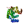
| ||||||||
|---|---|---|---|---|---|---|---|---|---|
| 1 |
| ||||||||
| Unit cell |
|
-Components
| #1: Protein | Mass: 24537.207 Da / Num. of mol.: 1 Source method: isolated from a genetically manipulated source Source: (gene. exp.) Homo sapiens (human) / Gene: PIMT_HUMAN / Organ: FETAL BRAIN / Plasmid: PET21C / Production host:  References: UniProt: P22061, protein-L-isoaspartate(D-aspartate) O-methyltransferase |
|---|---|
| #2: Chemical | ChemComp-SAH /   Type: L-peptide linking / Mass: 384.411 Da / Num. of mol.: 1 / Source method: obtained synthetically / Formula: C14H20N6O5S Type: L-peptide linking / Mass: 384.411 Da / Num. of mol.: 1 / Source method: obtained synthetically / Formula: C14H20N6O5S |
| #3: Water | ChemComp-HOH /  Mass: 18.015 Da / Num. of mol.: 279 / Source method: isolated from a natural source / Formula: H2O Mass: 18.015 Da / Num. of mol.: 279 / Source method: isolated from a natural source / Formula: H2O |
-Experimental details
-Experiment
| Experiment | Method: X-RAY DIFFRACTION / Number of used crystals: 1 |
|---|
- Sample preparation
| Crystal | Density Matthews: 2.31 Å3/Da / Density % sol: 46.86 % | ||||||||||||||||||||||||||||||||||||
|---|---|---|---|---|---|---|---|---|---|---|---|---|---|---|---|---|---|---|---|---|---|---|---|---|---|---|---|---|---|---|---|---|---|---|---|---|---|
| Crystal grow | Temperature: 277 K / Method: vapor diffusion, hanging drop / pH: 6.5 Details: PEG 8000, sodium cacodylate, magnesium acetate, DTT, bis-Tris acetate, S-adenosyl homocysteine, pH 6.5, VAPOR DIFFUSION, HANGING DROP, temperature 277K | ||||||||||||||||||||||||||||||||||||
| Crystal grow | *PLUS Temperature: 4 ℃ / Details: used macroseeding | ||||||||||||||||||||||||||||||||||||
| Components of the solutions | *PLUS
|
-Data collection
| Diffraction | Mean temperature: 93 K |
|---|---|
| Diffraction source | Source: ROTATING ANODE / Type: RIGAKU RU300 / Wavelength: 1.5418 |
| Detector | Type: RIGAKU RAXIS IV / Detector: IMAGE PLATE / Date: Nov 25, 1997 / Details: MSC MIRRORS |
| Radiation | Monochromator: NICKEL FILTER / Protocol: SINGLE WAVELENGTH / Monochromatic (M) / Laue (L): M / Scattering type: x-ray |
| Radiation wavelength | Wavelength: 1.5418 Å / Relative weight: 1 |
| Reflection | Resolution: 1.5→50 Å / Num. obs: 31332 / % possible obs: 86.8 % / Observed criterion σ(I): -3 / Redundancy: 3.47 % / Biso Wilson estimate: 18.1 Å2 / Rmerge(I) obs: 0.04 / Net I/σ(I): 28 |
| Reflection shell | Resolution: 1.5→1.55 Å / Redundancy: 1.16 % / Rmerge(I) obs: 0.205 / % possible all: 34.1 |
| Reflection | *PLUS Highest resolution: 1.6 Å / Lowest resolution: 50 Å / Num. obs: 28488 / % possible obs: 95.7 % / Redundancy: 1.7 % / Rmerge(I) obs: 0.039 |
| Reflection shell | *PLUS Highest resolution: 1.6 Å / Lowest resolution: 1.66 Å / % possible obs: 87.1 % / Redundancy: 1.4 % / Num. unique obs: 2587 / Rmerge(I) obs: 0.124 |
- Processing
| Software |
| ||||||||||||||||||||||||||||||||||||||||||||||||||||||||||||||||||||||||||||||||
|---|---|---|---|---|---|---|---|---|---|---|---|---|---|---|---|---|---|---|---|---|---|---|---|---|---|---|---|---|---|---|---|---|---|---|---|---|---|---|---|---|---|---|---|---|---|---|---|---|---|---|---|---|---|---|---|---|---|---|---|---|---|---|---|---|---|---|---|---|---|---|---|---|---|---|---|---|---|---|---|---|---|
| Refinement | Method to determine structure: MOLECULAR REPLACEMENT Starting model: 1DL5.PDB Resolution: 1.5→14.98 Å / Rfactor Rfree error: 0.005 / Data cutoff high absF: 944740.24 / Data cutoff high rms absF: 944740.24 / Data cutoff low absF: 0 / Isotropic thermal model: RESTRAINED / Cross valid method: THROUGHOUT / σ(F): 0 Details: BULK SOLVENT MODEL USED. THE SAH TOPOLOGY/PARAMETER FILES FOR CNS WERE CREATED BY COMBINING THE STANDARD MET AND ADE ENTRIES. THE DEFAULT CNS DNA-RNA_REP.PARAM FOR RNA/DNA WAS MODIFIED IN ...Details: BULK SOLVENT MODEL USED. THE SAH TOPOLOGY/PARAMETER FILES FOR CNS WERE CREATED BY COMBINING THE STANDARD MET AND ADE ENTRIES. THE DEFAULT CNS DNA-RNA_REP.PARAM FOR RNA/DNA WAS MODIFIED IN THE LATER STAGES OF REFINEMENT. THE RIBOSE RING DIHEDRAL FORCE CONSTANTS WERE SET TO 0.0 TO ALLOW THE RIBOSE RING PUCKER TO FIT THE ELECTRON DENSITY.
| ||||||||||||||||||||||||||||||||||||||||||||||||||||||||||||||||||||||||||||||||
| Solvent computation | Solvent model: FLAT MODEL / Bsol: 56.88 Å2 / ksol: 0.402 e/Å3 | ||||||||||||||||||||||||||||||||||||||||||||||||||||||||||||||||||||||||||||||||
| Displacement parameters | Biso mean: 18.4 Å2
| ||||||||||||||||||||||||||||||||||||||||||||||||||||||||||||||||||||||||||||||||
| Refine analyze |
| ||||||||||||||||||||||||||||||||||||||||||||||||||||||||||||||||||||||||||||||||
| Refinement step | Cycle: LAST / Resolution: 1.5→14.98 Å
| ||||||||||||||||||||||||||||||||||||||||||||||||||||||||||||||||||||||||||||||||
| Refine LS restraints |
| ||||||||||||||||||||||||||||||||||||||||||||||||||||||||||||||||||||||||||||||||
| LS refinement shell | Resolution: 1.5→1.59 Å / Rfactor Rfree error: 0.032 / Total num. of bins used: 6
| ||||||||||||||||||||||||||||||||||||||||||||||||||||||||||||||||||||||||||||||||
| Xplor file |
| ||||||||||||||||||||||||||||||||||||||||||||||||||||||||||||||||||||||||||||||||
| Refinement | *PLUS Highest resolution: 1.6 Å / Lowest resolution: 15 Å / Num. reflection obs: 27830 / σ(F): 0 / Num. reflection Rfree: 1384 / % reflection Rfree: 5 % / Rfactor obs: 0.18 / Rfactor Rfree: 0.211 | ||||||||||||||||||||||||||||||||||||||||||||||||||||||||||||||||||||||||||||||||
| Solvent computation | *PLUS | ||||||||||||||||||||||||||||||||||||||||||||||||||||||||||||||||||||||||||||||||
| Displacement parameters | *PLUS Biso mean: 18.4 Å2 | ||||||||||||||||||||||||||||||||||||||||||||||||||||||||||||||||||||||||||||||||
| Refine LS restraints | *PLUS
| ||||||||||||||||||||||||||||||||||||||||||||||||||||||||||||||||||||||||||||||||
| LS refinement shell | *PLUS Rfactor Rfree: 0.343 / % reflection Rfree: 4.9 % / Rfactor Rwork: 0.304 |
