 Movie
Movie Controller

[English] 日本語
 Yorodumi
- PDB-4huj: Crystal structure of a hypothetical protein SMa0349 from Sinorhiz... -
+ Open data

- Basic information
| Entry | Database: PDB / ID: 4huj | ||||||
|---|---|---|---|---|---|---|---|
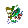
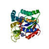
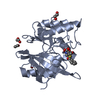
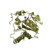
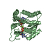
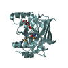
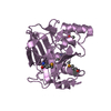
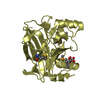
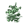
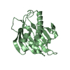
| Title | Crystal structure of a hypothetical protein SMa0349 from Sinorhizobium meliloti | ||||||
 Components Components | Uncharacterized protein | ||||||
 Keywords Keywords | STRUCTURAL GENOMICS / UNKNOWN FUNCTION / PSI-Biology / NYSGRC / New York Structural Genomics Research Consortium / dinucleotide-binding / NAD(P)+ binding | ||||||
| Function / homology | : / Pyrroline-5-carboxylate reductase, catalytic, N-terminal / NADP oxidoreductase coenzyme F420-dependent / NAD(P)-binding Rossmann-like Domain / NAD(P)-binding domain superfamily / Rossmann fold / 3-Layer(aba) Sandwich / Alpha Beta / Pyrroline-5-carboxylate reductase catalytic N-terminal domain-containing protein Function and homology information Function and homology information | ||||||
| Biological species |  Sinorhizobium meliloti (bacteria) Sinorhizobium meliloti (bacteria) | ||||||
| Method |  X-RAY DIFFRACTION / SYNCHROTRON / SAD / Resolution: 1.77 Å X-RAY DIFFRACTION / SYNCHROTRON / SAD / Resolution: 1.77 Å | ||||||
 Authors Authors | Rice, S. / Eswaramoorthy, S. / Chamala, S. / Evans, B. / Foti, F. / Gizzi, A. / Hillerich, B. / Kar, A. / LaFleur, J. / Seidel, R. ...Rice, S. / Eswaramoorthy, S. / Chamala, S. / Evans, B. / Foti, F. / Gizzi, A. / Hillerich, B. / Kar, A. / LaFleur, J. / Seidel, R. / Villigas, G. / Zencheck, W. / Almo, S.C. / Swaminathan, S. / New York Structural Genomics Research Consortium (NYSGRC) | ||||||
 Citation Citation | Journal: To be Published Title: Crystal structure of a hypothetical protein SMa0349 from Sinorhizobium meliloti Authors: Rice, S. / Eswaramoorthy, S. / Almo, S.C. / Swaminathan, S. | ||||||
| History |
|
- Structure visualization
| Structure viewer | Molecule: MolmilJmol/JSmol |
|---|
- Downloads & links
-Download
| PDBx/mmCIF format | 4huj.cif.gz | 89.3 KB | Display | PDBx/mmCIF format |
|---|---|---|---|---|
| PDB format | pdb4huj.ent.gz | 67.6 KB | Display | PDB format |
| PDBx/mmJSON format | 4huj.json.gz | Tree view | PDBx/mmJSON format | |
| Others |  Other downloads Other downloads |
-Validation report
| Arichive directory | https://data.pdbj.org/pub/pdb/validation_reports/hu/4hujftp://data.pdbj.org/pub/pdb/validation_reports/hu/4huj | HTTPS FTP |
|---|
-Related structure data
| Similar structure data | |
|---|---|
| Other databases |
-Links
 PDBj
PDBj- Assembly
| Deposited unit | 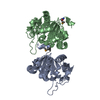
| ||||||||
|---|---|---|---|---|---|---|---|---|---|
| 1 | 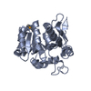
| ||||||||
| 2 | 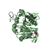
| ||||||||
| 3 | 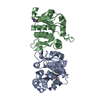
| ||||||||
| Unit cell |
|
-Components
| #1: Protein | Mass: 22992.660 Da / Num. of mol.: 2 Source method: isolated from a genetically manipulated source Source: (gene. exp.) Sinorhizobium meliloti (bacteria) / Strain: 1021 / Gene: RA0188, SMa0349 / Plasmid: PET3A / Production host: #2: Water | ChemComp-HOH / |  Mass: 18.015 Da / Num. of mol.: 178 / Source method: isolated from a natural source / Formula: H2O Mass: 18.015 Da / Num. of mol.: 178 / Source method: isolated from a natural source / Formula: H2OHas protein modification | Y | |
|---|
-Experimental details
-Experiment
| Experiment | Method: X-RAY DIFFRACTION / Number of used crystals: 1 |
|---|
- Sample preparation
| Crystal | Density Matthews: 2.53 Å3/Da / Density % sol: 51.37 % |
|---|---|
| Crystal grow | Temperature: 293 K / Method: vapor diffusion, sitting drop / pH: 8.5 Details: 0.2M Lithium sulfate, 0.1M TRIS, 30% PEG4000 and tert-Butanol, pH 8.5, VAPOR DIFFUSION, SITTING DROP, temperature 293K |
-Data collection
| Diffraction | Mean temperature: 100 K |
|---|---|
| Diffraction source | Source: SYNCHROTRON / Site: NSLS  / Beamline: X29A / Wavelength: 0.9792 Å / Beamline: X29A / Wavelength: 0.9792 Å |
| Detector | Type: ADSC QUANTUM 315 / Detector: CCD / Date: Jul 16, 2012 |
| Radiation | Protocol: SINGLE WAVELENGTH / Monochromatic (M) / Laue (L): M / Scattering type: x-ray |
| Radiation wavelength | Wavelength: 0.9792 Å / Relative weight: 1 |
| Reflection | Resolution: 1.77→50 Å / Num. all: 46300 / Num. obs: 46300 / % possible obs: 99.8 % / Observed criterion σ(F): 0 / Observed criterion σ(I): 0 / Redundancy: 14.2 % / Biso Wilson estimate: 19.2 Å2 / Rmerge(I) obs: 0.063 / Net I/σ(I): 16.4 |
| Reflection shell | Resolution: 1.77→1.82 Å / Redundancy: 13.7 % / Rmerge(I) obs: 0.335 / Num. unique all: 3763 / % possible all: 99.8 |
- Processing
| Software |
| |||||||||||||||||||||||||||||||||||||||||||||||||||||||||||||||||
|---|---|---|---|---|---|---|---|---|---|---|---|---|---|---|---|---|---|---|---|---|---|---|---|---|---|---|---|---|---|---|---|---|---|---|---|---|---|---|---|---|---|---|---|---|---|---|---|---|---|---|---|---|---|---|---|---|---|---|---|---|---|---|---|---|---|---|
| Refinement | Method to determine structure: SAD / Resolution: 1.77→42.614 Å / Cor.coef. Fo:Fc: 0.948 / Cor.coef. Fo:Fc free: 0.938 / SU B: 2.044 / SU ML: 0.067 / Cross valid method: THROUGHOUT / ESU R: 0.118 / ESU R Free: 0.113 / Stereochemistry target values: MAXIMUM LIKELIHOOD / Details: HYDROGENS HAVE BEEN ADDED IN THE RIDING POSITIONS
| |||||||||||||||||||||||||||||||||||||||||||||||||||||||||||||||||
| Solvent computation | Ion probe radii: 0.8 Å / Shrinkage radii: 0.8 Å / VDW probe radii: 1.4 Å / Solvent model: MASK | |||||||||||||||||||||||||||||||||||||||||||||||||||||||||||||||||
| Displacement parameters | Biso mean: 18.322 Å2
| |||||||||||||||||||||||||||||||||||||||||||||||||||||||||||||||||
| Refinement step | Cycle: LAST / Resolution: 1.77→42.614 Å
| |||||||||||||||||||||||||||||||||||||||||||||||||||||||||||||||||
| Refine LS restraints |
| |||||||||||||||||||||||||||||||||||||||||||||||||||||||||||||||||
| LS refinement shell | Resolution: 1.767→1.812 Å / Total num. of bins used: 20
|
