 Movie
Movie Controller

[English] 日本語
 Yorodumi
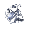
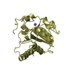
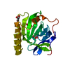
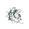
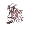
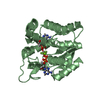
- PDB-1fvg: CRYSTAL STRUCTURE OF BOVINE PEPTIDE METHIONINE SULFOXIDE REDUCTASE -
+ Open data

- Basic information
| Entry | Database: PDB / ID: 1fvg | ||||||
|---|---|---|---|---|---|---|---|
| Title | CRYSTAL STRUCTURE OF BOVINE PEPTIDE METHIONINE SULFOXIDE REDUCTASE | ||||||
 Components Components | PEPTIDE METHIONINE SULFOXIDE REDUCTASE | ||||||
 Keywords Keywords | OXIDOREDUCTASE | ||||||
| Function / homology |  Function and homology information Function and homology informationProtein repair / L-methionine (S)-S-oxide reductase activity / peptide-methionine (S)-S-oxide reductase / peptide-methionine (S)-S-oxide reductase activity / cellular response to oxidative stress / mitochondrion / cytoplasm Similarity search - Function | ||||||
| Biological species |  | ||||||
| Method |  X-RAY DIFFRACTION / SYNCHROTRON / MAD / Resolution: 1.6 Å X-RAY DIFFRACTION / SYNCHROTRON / MAD / Resolution: 1.6 Å | ||||||
 Authors Authors | Lowther, W.T. / Brot, N. / Weissbach, H. / Matthews, B.W. | ||||||
 Citation Citation | Journal: Biochemistry / Year: 2000 Title: Structure and mechanism of peptide methionine sulfoxide reductase, an "anti-oxidation" enzyme. Authors: Lowther, W.T. / Brot, N. / Weissbach, H. / Matthews, B.W. #1: Journal: Proc.Natl.Acad.Sci.USA / Year: 2000Title: Thiol-disulfide exchange is Involved in the Catalytic Mechanism of Peptide Methionine Sulfoxide Reductase Authors: Lowther, W.T. / Brot, N. / Weissbach, H. / Honek, J.F. / Matthews, B.W. | ||||||
| History |
|
- Structure visualization
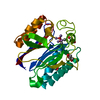
| Structure viewer | Molecule: MolmilJmol/JSmol |
|---|
- Downloads & links
-Download
| PDBx/mmCIF format | 1fvg.cif.gz | 55.4 KB | Display | PDBx/mmCIF format |
|---|---|---|---|---|
| PDB format | pdb1fvg.ent.gz | 38.9 KB | Display | PDB format |
| PDBx/mmJSON format | 1fvg.json.gz | Tree view | PDBx/mmJSON format | |
| Others |  Other downloads Other downloads |
-Validation report
| Arichive directory | https://data.pdbj.org/pub/pdb/validation_reports/fv/1fvgftp://data.pdbj.org/pub/pdb/validation_reports/fv/1fvg | HTTPS FTP |
|---|
-Related structure data


-Links
 PDBj
PDBj


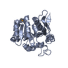
- Assembly
| Deposited unit | 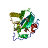
| ||||||||
|---|---|---|---|---|---|---|---|---|---|
| 1 |
| ||||||||
| Unit cell |
|
-Components
| #1: Protein | Mass: 22385.301 Da / Num. of mol.: 1 Source method: isolated from a genetically manipulated source Details: DITHIOTHREITOL COMPLEX / Source: (gene. exp.)  |
|---|---|
| #2: Chemical | ChemComp-DTT /   Mass: 154.251 Da / Num. of mol.: 1 / Source method: obtained synthetically / Formula: C4H10O2S2 Mass: 154.251 Da / Num. of mol.: 1 / Source method: obtained synthetically / Formula: C4H10O2S2 |
| #3: Water | ChemComp-HOH /  Mass: 18.015 Da / Num. of mol.: 140 / Source method: isolated from a natural source / Formula: H2O Mass: 18.015 Da / Num. of mol.: 140 / Source method: isolated from a natural source / Formula: H2O |
| Has protein modification | Y |
-Experimental details
-Experiment
| Experiment | Method: X-RAY DIFFRACTION / Number of used crystals: 1 |
|---|
- Sample preparation
| Crystal | Density Matthews: 2.1 Å3/Da / Density % sol: 41.55 % | |||||||||||||||||||||||||||||||||||||||||||||||||||||||||||||||
|---|---|---|---|---|---|---|---|---|---|---|---|---|---|---|---|---|---|---|---|---|---|---|---|---|---|---|---|---|---|---|---|---|---|---|---|---|---|---|---|---|---|---|---|---|---|---|---|---|---|---|---|---|---|---|---|---|---|---|---|---|---|---|---|---|
| Crystal grow | Temperature: 298 K / Method: vapor diffusion, hanging drop / pH: 4.7 Details: dithiothreitol, PEG 8000, citrate, Na2HPO4, dimethyl sulfoxide, NaCl, pH 4.70, VAPOR DIFFUSION, HANGING DROP, temperature 298K | |||||||||||||||||||||||||||||||||||||||||||||||||||||||||||||||
| Crystal grow | *PLUS pH: 7.4 / Method: vapor diffusion | |||||||||||||||||||||||||||||||||||||||||||||||||||||||||||||||
| Components of the solutions | *PLUS
|
-Data collection
| Diffraction | Mean temperature: 103 K | ||||||||||||
|---|---|---|---|---|---|---|---|---|---|---|---|---|---|
| Diffraction source | Source: SYNCHROTRON / Site: SSRL  / Beamline: BL9-2 / Wavelength: 0.9787,0.9788,0.9537 / Beamline: BL9-2 / Wavelength: 0.9787,0.9788,0.9537 | ||||||||||||
| Detector | Type: ADSC QUANTUM 4 / Detector: CCD / Date: Apr 21, 2000 | ||||||||||||
| Radiation | Protocol: MAD / Monochromatic (M) / Laue (L): M / Scattering type: x-ray | ||||||||||||
| Radiation wavelength |
| ||||||||||||
| Reflection | Resolution: 1.6→53.5 Å / Num. all: 86018 / Num. obs: 23472 / % possible obs: 95.8 % / Observed criterion σ(I): 8 / Redundancy: 4 % / Biso Wilson estimate: 15.9 Å2 / Rmerge(I) obs: 0.038 / Net I/σ(I): 37.2 | ||||||||||||
| Reflection shell | Resolution: 1.6→1.66 Å / Redundancy: 4 % / Rmerge(I) obs: 0.171 / % possible all: 93.9 | ||||||||||||
| Reflection | *PLUS Num. measured all: 86018 | ||||||||||||
| Reflection shell | *PLUS % possible obs: 93.9 % / Mean I/σ(I) obs: 8 |
- Processing
| Software |
| ||||||||||||||||||||||||||||||||||||||||
|---|---|---|---|---|---|---|---|---|---|---|---|---|---|---|---|---|---|---|---|---|---|---|---|---|---|---|---|---|---|---|---|---|---|---|---|---|---|---|---|---|---|
| Refinement | Method to determine structure: MAD / Resolution: 1.6→53.58 Å / σ(I): 8 / Stereochemistry target values: Engh & Huber Details: The structure factors represent the "optimized" reference structure factors (Fpsha) from a three wavelength MAD experiment analyzed by SHARP.
| ||||||||||||||||||||||||||||||||||||||||
| Solvent computation | Bsol: 141.2 Å2 / ksol: 0.802 e/Å3 | ||||||||||||||||||||||||||||||||||||||||
| Refinement step | Cycle: LAST / Resolution: 1.6→53.58 Å
| ||||||||||||||||||||||||||||||||||||||||
| Refine LS restraints |
| ||||||||||||||||||||||||||||||||||||||||
| Software | *PLUS Name: TNT / Version: 5E / Classification: refinement | ||||||||||||||||||||||||||||||||||||||||
| Refinement | *PLUS Highest resolution: 1.6 Å / % reflection Rfree: 10 % / Rfactor obs: 0.182 | ||||||||||||||||||||||||||||||||||||||||
| Solvent computation | *PLUS | ||||||||||||||||||||||||||||||||||||||||
| Displacement parameters | *PLUS | ||||||||||||||||||||||||||||||||||||||||
| Refine LS restraints | *PLUS
|
