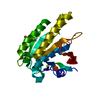
[English] 日本語
Yorodumi
- PDB-5zdm: The ligand-free structure of FomD -

+
Open data


ID or keywords:

Loading...

-
Basic information

Entry
Database: PDB / ID: 5zdm
TitleThe ligand-free structure of FomD
ComponentsFomD
KeywordsHYDROLASE / metalloprotein
Function / homologyDomain of unknown function DUF402 / FomD-like superfamily / Protein of unknown function (DUF402) / FomD
Function and homology information
Biological speciesStreptomyces fradiae (bacteria)
MethodX-RAY DIFFRACTION / SYNCHROTRON / MOLECULAR REPLACEMENT / Resolution: 1.38 Å
AuthorsSato, S. / Miyanaga, A. / Kudo, F. / Eguchi, T.
CitationJournal: Biochemistry / Year: 2018
Title: Biochemical and Structural Analysis of FomD That Catalyzes the Hydrolysis of Cytidylyl ( S)-2-Hydroxypropylphosphonate in Fosfomycin Biosynthesis.
Authors: Sato, S. / Miyanaga, A. / Kim, S.Y. / Kuzuyama, T. / Kudo, F. / Eguchi, T.
History
DepositionFeb 23, 2018Deposition site: PDBJ / Processing site: PDBJ
Revision 1.0Jul 25, 2018Provider: repository / Type: Initial release
Revision 1.1Aug 29, 2018Group: Data collection / Database references / Category: citation / citation_author
Item: _citation.journal_id_ISSN / _citation.journal_volume ..._citation.journal_id_ISSN / _citation.journal_volume / _citation.page_first / _citation.page_last / _citation.pdbx_database_id_PubMed / _citation.title / _citation_author.identifier_ORCID
Revision 1.2Nov 22, 2023Group: Data collection / Database references ...Data collection / Database references / Derived calculations / Refinement description
Category: chem_comp_atom / chem_comp_bond ...chem_comp_atom / chem_comp_bond / database_2 / pdbx_initial_refinement_model / pdbx_struct_conn_angle / struct_conn
Item: _database_2.pdbx_DOI / _database_2.pdbx_database_accession ..._database_2.pdbx_DOI / _database_2.pdbx_database_accession / _pdbx_struct_conn_angle.ptnr1_auth_comp_id / _pdbx_struct_conn_angle.ptnr1_auth_seq_id / _pdbx_struct_conn_angle.ptnr1_label_asym_id / _pdbx_struct_conn_angle.ptnr1_label_atom_id / _pdbx_struct_conn_angle.ptnr1_label_comp_id / _pdbx_struct_conn_angle.ptnr1_label_seq_id / _pdbx_struct_conn_angle.ptnr1_symmetry / _pdbx_struct_conn_angle.ptnr3_auth_comp_id / _pdbx_struct_conn_angle.ptnr3_auth_seq_id / _pdbx_struct_conn_angle.ptnr3_label_asym_id / _pdbx_struct_conn_angle.ptnr3_label_atom_id / _pdbx_struct_conn_angle.ptnr3_label_comp_id / _pdbx_struct_conn_angle.ptnr3_label_seq_id / _pdbx_struct_conn_angle.ptnr3_symmetry / _pdbx_struct_conn_angle.value / _struct_conn.pdbx_dist_value / _struct_conn.ptnr1_auth_comp_id / _struct_conn.ptnr1_auth_seq_id / _struct_conn.ptnr1_label_asym_id / _struct_conn.ptnr1_label_atom_id / _struct_conn.ptnr1_label_comp_id / _struct_conn.ptnr1_label_seq_id / _struct_conn.ptnr2_auth_comp_id / _struct_conn.ptnr2_auth_seq_id / _struct_conn.ptnr2_label_asym_id / _struct_conn.ptnr2_label_atom_id / _struct_conn.ptnr2_label_comp_id / _struct_conn.ptnr2_symmetry

-
Structure visualization

Structure viewerMolecule:
MolmilJmol/JSmol

Downloads & links

-
Assembly

Deposited unit
A: FomD
hetero molecules


Theoretical massNumber of molelcules
Total (without water)24,3526
Polymers23,9951
Non-polymers3565
Water3,783210
1


  • Idetical with deposited unit
  • defined by author
  • Evidence: gel filtration
TypeNameSymmetry operationNumber
identity operation1_555x,y,z1
Buried area800 Å2
ΔGint-18 kcal/mol
Surface area10170 Å2
Unit cell
Length a, b, c (Å)40.474, 46.215, 52.504
Angle α, β, γ (deg.)90.00, 110.27, 90.00
Int Tables number4
Space group name H-MP1211

-
Components

#1: Protein FomD


Mass: 23995.363 Da / Num. of mol.: 1 / Mutation: L139F
Source method: isolated from a genetically manipulated source
Source: (gene. exp.) Streptomyces fradiae (bacteria) / Gene: fomD / Plasmid: pHis8 / Production host: Escherichia coli BL21(DE3) (bacteria) / Strain (production host): BL21(DE3) / References: UniProt: D2SNF7
#2: Chemical ChemComp-CA / CALCIUM ION


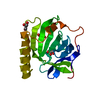
Mass: 40.078 Da / Num. of mol.: 2 / Source method: obtained synthetically / Formula: Ca
#3: Chemical ChemComp-GOL / GLYCEROL / GLYCERIN / PROPANE-1,2,3-TRIOL


Mass: 92.094 Da / Num. of mol.: 3 / Source method: obtained synthetically / Formula: C3H8O3
#4: Water ChemComp-HOH / water


Mass: 18.015 Da / Num. of mol.: 210 / Source method: isolated from a natural source / Formula: H2O

-
Experimental details

-
Experiment

ExperimentMethod: X-RAY DIFFRACTION / Number of used crystals: 1

-
Sample preparation

CrystalDensity Matthews: 1.92 Å3/Da / Density % sol: 35.93 %
Crystal growTemperature: 278 K / Method: vapor diffusion, sitting drop / pH: 8.5
Details: Tris-HCl, calcium chloride, PEG 8000, trimethylamine N-oxide, cytosine

-
Data collection

DiffractionMean temperature: 100 K
Diffraction sourceSource: SYNCHROTRON / Site: Photon Factory / Beamline: AR-NW12A / Wavelength: 1 Å
DetectorType: ADSC QUANTUM 270 / Detector: CCD / Date: Dec 22, 2017
RadiationMonochromator: Numerical link type Si(111) double crystal monochromator
Protocol: SINGLE WAVELENGTH / Monochromatic (M) / Laue (L): M / Scattering type: x-ray
Radiation wavelengthWavelength: 1 Å / Relative weight: 1
ReflectionResolution: 1.38→50 Å / Num. obs: 36195 / % possible obs: 96.4 % / Redundancy: 3.6 % / Rmerge(I) obs: 0.031 / Net I/σ(I): 18
Reflection shellResolution: 1.38→1.41 Å / Redundancy: 3.2 % / Rmerge(I) obs: 0.399 / Mean I/σ(I) obs: 2.4 / Num. unique obs: 1840 / % possible all: 94.3

-
Processing

Software
NameVersionClassification
REFMAC5.7.0029refinement
XDSdata reduction
XDSdata scaling
MOLREPphasing
ARP/wARPmodel building
RefinementMethod to determine structure: MOLECULAR REPLACEMENT
Starting model: 3EXM
Resolution: 1.38→49.25 Å / Cor.coef. Fo:Fc: 0.971 / Cor.coef. Fo:Fc free: 0.959 / SU B: 1.97 / SU ML: 0.04 / Cross valid method: THROUGHOUT / ESU R: 0.061 / ESU R Free: 0.064 / Details: HYDROGENS HAVE BEEN ADDED IN THE RIDING POSITIONS
RfactorNum. reflection% reflectionSelection details
Rfree0.19178 1756 4.9 %RANDOM
Rwork0.1635 ---
obs0.16486 34427 96.39 %-
Solvent computationIon probe radii: 0.8 Å / Shrinkage radii: 0.8 Å / VDW probe radii: 1.2 Å
Displacement parametersBiso mean: 20.711 Å2
Baniso -1Baniso -2Baniso -3
1-0.81 Å2-0 Å20.42 Å2
2---1.38 Å20 Å2
3---0.33 Å2
Refinement stepCycle: 1 / Resolution: 1.38→49.25 Å
ProteinNucleic acidLigandSolventTotal
Num. atoms1619 0 20 210 1849
Refine LS restraints
Refine-IDTypeDev idealDev ideal targetNumber
X-RAY DIFFRACTIONr_bond_refined_d0.0260.0191724
X-RAY DIFFRACTIONr_bond_other_d0.0020.021535
X-RAY DIFFRACTIONr_angle_refined_deg2.3841.9042354
X-RAY DIFFRACTIONr_angle_other_deg1.02833512
X-RAY DIFFRACTIONr_dihedral_angle_1_deg6.5895203
X-RAY DIFFRACTIONr_dihedral_angle_2_deg35.75122.10595
X-RAY DIFFRACTIONr_dihedral_angle_3_deg13.46915244
X-RAY DIFFRACTIONr_dihedral_angle_4_deg18.2761521
X-RAY DIFFRACTIONr_chiral_restr0.140.2233
X-RAY DIFFRACTIONr_gen_planes_refined0.0160.0211986
X-RAY DIFFRACTIONr_gen_planes_other0.0020.02461
X-RAY DIFFRACTIONr_nbd_refined
X-RAY DIFFRACTIONr_nbd_other
X-RAY DIFFRACTIONr_nbtor_refined
X-RAY DIFFRACTIONr_nbtor_other
X-RAY DIFFRACTIONr_xyhbond_nbd_refined
X-RAY DIFFRACTIONr_xyhbond_nbd_other
X-RAY DIFFRACTIONr_metal_ion_refined
X-RAY DIFFRACTIONr_metal_ion_other
X-RAY DIFFRACTIONr_symmetry_vdw_refined
X-RAY DIFFRACTIONr_symmetry_vdw_other
X-RAY DIFFRACTIONr_symmetry_hbond_refined
X-RAY DIFFRACTIONr_symmetry_hbond_other
X-RAY DIFFRACTIONr_symmetry_metal_ion_refined
X-RAY DIFFRACTIONr_symmetry_metal_ion_other
X-RAY DIFFRACTIONr_mcbond_it
X-RAY DIFFRACTIONr_mcbond_other
X-RAY DIFFRACTIONr_mcangle_it
X-RAY DIFFRACTIONr_mcangle_other
X-RAY DIFFRACTIONr_scbond_it
X-RAY DIFFRACTIONr_scbond_other
X-RAY DIFFRACTIONr_scangle_it
X-RAY DIFFRACTIONr_scangle_other
X-RAY DIFFRACTIONr_long_range_B_refined
X-RAY DIFFRACTIONr_long_range_B_other
X-RAY DIFFRACTIONr_rigid_bond_restr
X-RAY DIFFRACTIONr_sphericity_free
X-RAY DIFFRACTIONr_sphericity_bonded
LS refinement shellResolution: 1.38→1.416 Å / Total num. of bins used: 20
RfactorNum. reflection% reflection
Rfree0.249 125 -
Rwork0.249 2512 -
obs--95.47 %
Refinement TLS params.Method: refined / Origin x: 8.848 Å / Origin y: -0.599 Å / Origin z: 9.216 Å
111213212223313233
T0.0527 Å20.0041 Å20.0058 Å2-0.1219 Å2-0.0083 Å2--0.0038 Å2
L0.3163 °2-0.2193 °2-0.1859 °2-0.5846 °20.0466 °2--0.6243 °2
S0.0257 Å °0.0136 Å °-0.0065 Å °-0.0428 Å °-0.0578 Å °0.0256 Å °0.0349 Å °-0.0459 Å °0.0321 Å °
Refinement TLS group
IDRefine-IDRefine TLS-IDAuth asym-IDAuth seq-ID
1X-RAY DIFFRACTION1A10 - 207
2X-RAY DIFFRACTION1A301 - 305

+
About Yorodumi

-
News

-
Feb 9, 2022. New format data for meta-information of EMDB entries

New format data for meta-information of EMDB entries

  • Version 3 of the EMDB header file is now the official format.
  • The previous official version 1.9 will be removed from the archive.

Related info.:EMDB header

External links:wwPDB to switch to version 3 of the EMDB data model

-
Aug 12, 2020. Covid-19 info

Covid-19 info

URL: https://pdbjlvh1.pdbj.org/emnavi/covid19.php

New page: Covid-19 featured information page in EM Navigator.

Related info.:Covid-19 info / Mar 5, 2020. Novel coronavirus structure data

+
Mar 5, 2020. Novel coronavirus structure data

Novel coronavirus structure data

Related info.:Yorodumi Speices / Aug 12, 2020. Covid-19 info

External links:COVID-19 featured content - PDBj / Molecule of the Month (242):Coronavirus Proteases

+
Jan 31, 2019. EMDB accession codes are about to change! (news from PDBe EMDB page)

EMDB accession codes are about to change! (news from PDBe EMDB page)

  • The allocation of 4 digits for EMDB accession codes will soon come to an end. Whilst these codes will remain in use, new EMDB accession codes will include an additional digit and will expand incrementally as the available range of codes is exhausted. The current 4-digit format prefixed with “EMD-” (i.e. EMD-XXXX) will advance to a 5-digit format (i.e. EMD-XXXXX), and so on. It is currently estimated that the 4-digit codes will be depleted around Spring 2019, at which point the 5-digit format will come into force.
  • The EM Navigator/Yorodumi systems omit the EMD- prefix.

Related info.:Q: What is EMD? / ID/Accession-code notation in Yorodumi/EM Navigator

External links:EMDB Accession Codes are Changing Soon! / Contact to PDBj

+
Jul 12, 2017. Major update of PDB

Major update of PDB

  • wwPDB released updated PDB data conforming to the new PDBx/mmCIF dictionary.
  • This is a major update changing the version number from 4 to 5, and with Remediation, in which all the entries are updated.
  • In this update, many items about electron microscopy experimental information are reorganized (e.g. em_software).
  • Now, EM Navigator and Yorodumi are based on the updated data.

External links:wwPDB Remediation / Enriched Model Files Conforming to OneDep Data Standards Now Available in the PDB FTP Archive

-
Yorodumi

Thousand views of thousand structures

  • Yorodumi is a browser for structure data from EMDB, PDB, SASBDB, etc.
  • This page is also the successor to EM Navigator detail page, and also detail information page/front-end page for Omokage search.
  • The word "yorodu" (or yorozu) is an old Japanese word meaning "ten thousand". "mi" (miru) is to see.

Related info.:EMDB / PDB / SASBDB / Comparison of 3 databanks / Yorodumi Search / Aug 31, 2016. New EM Navigator & Yorodumi / Yorodumi Papers / Jmol/JSmol / Function and homology information / Changes in new EM Navigator and Yorodumi

Read more