 Movie
Movie Controller

[English] 日本語
 Yorodumi
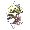
- PDB-3kkf: Crystal structure of Putative antibiotic biosynthesis monooxygena... -
+ Open data

- Basic information
| Entry | Database: PDB / ID: 3kkf | ||||||
|---|---|---|---|---|---|---|---|
| Title | Crystal structure of Putative antibiotic biosynthesis monooxygenase (NP_810307.1) from Bacteriodes thetaiotaomicron VPI-5482 at 1.30 A resolution | ||||||
 Components Components | Putative antibiotic biosynthesis monooxygenase | ||||||
 Keywords Keywords | OXIDOREDUCTASE / Putative antibiotic biosynthesis monooxygenase / Structural Genomics / Joint Center for Structural Genomics / JCSG / Protein Structure Initiative / PSI-2 | ||||||
| Function / homology |  Function and homology information Function and homology information | ||||||
| Biological species |  Bacteroides thetaiotaomicron (bacteria) Bacteroides thetaiotaomicron (bacteria) | ||||||
| Method |  X-RAY DIFFRACTION / SYNCHROTRON / SAD / Resolution: 1.3 Å X-RAY DIFFRACTION / SYNCHROTRON / SAD / Resolution: 1.3 Å | ||||||
 Authors Authors | Joint Center for Structural Genomics (JCSG) | ||||||
 Citation Citation | Journal: To be published Title: Crystal structure of Putative antibiotic biosynthesis monooxygenase (NP_810307.1) from Bacteriodes thetaiotaomicron VPI-5482 at 1.30 A resolution Authors: Joint Center for Structural Genomics (JCSG) | ||||||
| History |
|
- Structure visualization
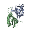
| Structure viewer | Molecule: MolmilJmol/JSmol |
|---|
- Downloads & links
-Download
| PDBx/mmCIF format | 3kkf.cif.gz | 72.1 KB | Display | PDBx/mmCIF format |
|---|---|---|---|---|
| PDB format | pdb3kkf.ent.gz | 53.2 KB | Display | PDB format |
| PDBx/mmJSON format | 3kkf.json.gz | Tree view | PDBx/mmJSON format | |
| Others |  Other downloads Other downloads |
-Validation report
| Arichive directory | https://data.pdbj.org/pub/pdb/validation_reports/kk/3kkfftp://data.pdbj.org/pub/pdb/validation_reports/kk/3kkf | HTTPS FTP |
|---|
-Related structure data
| Similar structure data | |
|---|---|
| Other databases |


-Links
 PDBj
PDBj- Assembly
| Deposited unit | 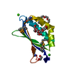
| ||||||||||||||||||
|---|---|---|---|---|---|---|---|---|---|---|---|---|---|---|---|---|---|---|---|
| 1 | 
| ||||||||||||||||||
| 2 | x 6
| ||||||||||||||||||
| Unit cell |
| ||||||||||||||||||
| Components on special symmetry positions |
|
-Components
-Protein , 1 types, 1 molecules A
| #1: Protein | Mass: 12307.491 Da / Num. of mol.: 1 / Fragment: sequence database residues 28-131 Source method: isolated from a genetically manipulated source Source: (gene. exp.) Bacteroides thetaiotaomicron (bacteria)Gene: BT_1394 / Plasmid: SpeedET / Production host: |
|---|
-Non-polymers , 5 types, 145 molecules 


| #2: Chemical |  Mass: 40.078 Da / Num. of mol.: 2 / Source method: obtained synthetically / Formula: Ca Mass: 40.078 Da / Num. of mol.: 2 / Source method: obtained synthetically / Formula: Ca#3: Chemical | ChemComp-ACT / |  Mass: 59.044 Da / Num. of mol.: 1 / Source method: obtained synthetically / Formula: C2H3O2 Mass: 59.044 Da / Num. of mol.: 1 / Source method: obtained synthetically / Formula: C2H3O2#4: Chemical | ChemComp-EDO / |  Mass: 62.068 Da / Num. of mol.: 1 / Source method: obtained synthetically / Formula: C2H6O2 Mass: 62.068 Da / Num. of mol.: 1 / Source method: obtained synthetically / Formula: C2H6O2#5: Chemical |  Mass: 282.331 Da / Num. of mol.: 2 / Source method: obtained synthetically / Formula: C12H26O7 / Comment: precipitant*YM Mass: 282.331 Da / Num. of mol.: 2 / Source method: obtained synthetically / Formula: C12H26O7 / Comment: precipitant*YM#6: Water | ChemComp-HOH / | Mass: 18.015 Da / Num. of mol.: 139 / Source method: isolated from a natural source / Formula: H2O |
|---|
-Details
| Has protein modification | Y |
|---|---|
| Sequence details | THIS CONSTRUCT (28-131) WAS EXPRESSED WITH THE PURIFICATION TAG MGSDKIHHHHHHENLYFQG. THE TAG WAS ...THIS CONSTRUCT (28-131) WAS EXPRESSED WITH THE PURIFICATI |
-Experimental details
-Experiment
| Experiment | Method: X-RAY DIFFRACTION / Number of used crystals: 1 |
|---|
- Sample preparation
| Crystal | Density Matthews: 1.88 Å3/Da / Density % sol: 34.52 % |
|---|---|
| Crystal grow | Temperature: 277 K / Method: vapor diffusion, sitting drop / pH: 7.5 Details: 0.2000M Ca(OAc)2, 40.0000% PEG-400, 0.1M HEPES pH 7.5, NANODROP, VAPOR DIFFUSION, SITTING DROP, temperature 277K |
-Data collection
| Diffraction | Mean temperature: 100 K | ||||||||||||||||||||||||||||||||||||||||||||||||||||||||||||||||||
|---|---|---|---|---|---|---|---|---|---|---|---|---|---|---|---|---|---|---|---|---|---|---|---|---|---|---|---|---|---|---|---|---|---|---|---|---|---|---|---|---|---|---|---|---|---|---|---|---|---|---|---|---|---|---|---|---|---|---|---|---|---|---|---|---|---|---|---|
| Diffraction source | Source: SYNCHROTRON / Site: SSRL  / Beamline: BL9-2 / Wavelength: 0.97919 / Beamline: BL9-2 / Wavelength: 0.97919 | ||||||||||||||||||||||||||||||||||||||||||||||||||||||||||||||||||
| Detector | Type: MARMOSAIC 325 mm CCD / Detector: CCD / Date: May 15, 2009 / Details: Flat collimating mirror, toroid focusing mirror | ||||||||||||||||||||||||||||||||||||||||||||||||||||||||||||||||||
| Radiation | Monochromator: Double crystal monochromator / Protocol: SINGLE WAVELENGTH / Monochromatic (M) / Laue (L): M / Scattering type: x-ray | ||||||||||||||||||||||||||||||||||||||||||||||||||||||||||||||||||
| Radiation wavelength | Wavelength: 0.97919 Å / Relative weight: 1 | ||||||||||||||||||||||||||||||||||||||||||||||||||||||||||||||||||
| Reflection | Resolution: 1.3→29.185 Å / Num. obs: 22848 / % possible obs: 99.2 % / Observed criterion σ(I): -3 / Redundancy: 7.0562 % / Biso Wilson estimate: 13.027 Å2 / Rmerge(I) obs: 0.043 / Net I/σ(I): 16.03 | ||||||||||||||||||||||||||||||||||||||||||||||||||||||||||||||||||
| Reflection shell |
|
-Phasing
| Phasing | Method: SAD |
|---|
- Processing
| Software |
| ||||||||||||||||||||||||||||||||||||||||||||||||||||||||||||||||||||||||||||||||||||||||||||||||||||
|---|---|---|---|---|---|---|---|---|---|---|---|---|---|---|---|---|---|---|---|---|---|---|---|---|---|---|---|---|---|---|---|---|---|---|---|---|---|---|---|---|---|---|---|---|---|---|---|---|---|---|---|---|---|---|---|---|---|---|---|---|---|---|---|---|---|---|---|---|---|---|---|---|---|---|---|---|---|---|---|---|---|---|---|---|---|---|---|---|---|---|---|---|---|---|---|---|---|---|---|---|---|
| Refinement | Method to determine structure: SAD / Resolution: 1.3→29.185 Å / Cor.coef. Fo:Fc: 0.977 / Cor.coef. Fo:Fc free: 0.963 / Occupancy max: 1 / Occupancy min: 0.08 / SU B: 1.479 / SU ML: 0.028 / Cross valid method: THROUGHOUT / σ(F): 0 / ESU R: 0.057 / ESU R Free: 0.051 Stereochemistry target values: MAXIMUM LIKELIHOOD WITH PHASES Details: 1. HYDROGENS HAVE BEEN ADDED IN THE RIDING POSITIONS. 2. A MET-INHIBITION PROTOCOL WAS USED FOR SELENOMETHIONINE INCORPORATION DURING PROTEIN EXPRESSION. THE OCCUPANCY OF THE SE ATOMS IN THE ...Details: 1. HYDROGENS HAVE BEEN ADDED IN THE RIDING POSITIONS. 2. A MET-INHIBITION PROTOCOL WAS USED FOR SELENOMETHIONINE INCORPORATION DURING PROTEIN EXPRESSION. THE OCCUPANCY OF THE SE ATOMS IN THE MSE RESIDUES WAS REDUCED TO 0.75 TO ACCOUNT FOR THE REDUCED SCATTERING POWER DUE TO PARTIAL S-MET INCORPORATION. 3. CALCIUM IONS (CA) FROM THE CRYSTALLIZATION SOLUTIONS HAVE BEEN MODELED INTO THE STRUCTURE BASED ON METAL EXCITATION SCAN AND COORDINATION GEOMETRY. 4. ETHYLENE GLYCOL (EDO) AND POLYETHYLENE GLYCOL (PEG AND P6G) FROM THE CRYSTALLIZATION/CRYOPROTECTANT SOLUTIONS HAVE BEEN MODELED INTO THE SOLVENT STRUCTURE.
| ||||||||||||||||||||||||||||||||||||||||||||||||||||||||||||||||||||||||||||||||||||||||||||||||||||
| Solvent computation | Ion probe radii: 0.8 Å / Shrinkage radii: 0.8 Å / VDW probe radii: 1.4 Å / Solvent model: MASK | ||||||||||||||||||||||||||||||||||||||||||||||||||||||||||||||||||||||||||||||||||||||||||||||||||||
| Displacement parameters | Biso max: 55.74 Å2 / Biso mean: 15.901 Å2 / Biso min: 8.38 Å2
| ||||||||||||||||||||||||||||||||||||||||||||||||||||||||||||||||||||||||||||||||||||||||||||||||||||
| Refinement step | Cycle: LAST / Resolution: 1.3→29.185 Å
| ||||||||||||||||||||||||||||||||||||||||||||||||||||||||||||||||||||||||||||||||||||||||||||||||||||
| Refine LS restraints |
| ||||||||||||||||||||||||||||||||||||||||||||||||||||||||||||||||||||||||||||||||||||||||||||||||||||
| LS refinement shell | Resolution: 1.3→1.334 Å / Total num. of bins used: 20
|
