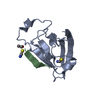
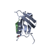
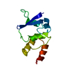
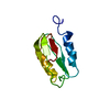
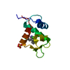
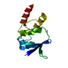
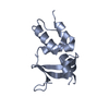
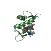
Entry Database : PDB / ID : 5ovpTitle PDZ domain from rat Shank3 bound to the C terminus of CIRL Adhesion G protein-coupled receptor L1 SH3 and multiple ankyrin repeat domains protein 3 Keywords / / / / Function / homology Function Domain/homology Component
/ / / / / / / / / / / / / / / / / / / / / / / / / / / / / / / / / / / / / / / / / / / / / / / / / / / / / / / / / / / / / / / / / / / / / / / / / / / / / / / / / / / / / / / / / / / / / / / / / / / / / / / / / / / / / / / / / / / / / / / / / / / / / / / / / / / / / / / / / / / / / / / / / / / / / / / / / / / Biological species Rattus norvegicus (Norway rat)Method / / Resolution : 1.5 Å Authors Ponna, S.K. / Myllykoski, M. / Boeckers, T.M. / Kursula, P. Journal : J. Neurochem. / Year : 2018Title : Structural basis for PDZ domain interactions in the post-synaptic density scaffolding protein Shank3.Authors : Ponna, S.K. / Ruskamo, S. / Myllykoski, M. / Keller, C. / Boeckers, T.M. / Kursula, P. History Deposition Aug 29, 2017 Deposition site / Processing site Revision 1.0 Mar 7, 2018 Provider / Type Revision 1.1 Jul 11, 2018 Group / Database references / Category Item / _citation.page_first / _citation.page_lastRevision 1.2 Oct 23, 2024 Group / Database references / Structure summaryCategory chem_comp_atom / chem_comp_bond ... chem_comp_atom / chem_comp_bond / database_2 / pdbx_entry_details / pdbx_modification_feature Item / _database_2.pdbx_database_accession
Show all Show less
 Movie
Movie Controller


 Open data
 Basic information
 Components
 Keywords
 Function and homology information
 X-RAY DIFFRACTION /
  Authors
 Citation
 Structure visualization
 Downloads & links
 Other downloads
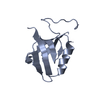


 PDBj
PDBj

 Assembly

 Mass: 18.015 Da / Num. of mol.: 63 / Source method: isolated from a natural source / Formula: H2O
 Sample preparation
 / Beamline: P13 (MX1) / Wavelength: 1 Å
 Processing