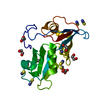
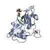
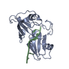
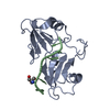
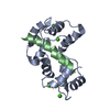
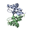
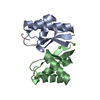
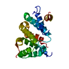
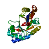
Entry Database : PDB / ID : 5o1wTitle Structure of Nrd1 RNA binding domain Protein NRD1 Keywords / / / / / Function / homology Function Domain/homology Component
/ / / / / / / / / / / / / / / / / / / / / / / / / / / / / / / / / / / / / / / / / / / / Biological species Saccharomyces cerevisiae (brewer's yeast)Method / / / Resolution : 2.3 Å Authors Franco-Echevarria, E. / Perez-Canadillas, J.M. / Gonzalez, B. Funding support Organization Grant number Country CSIC
Journal : Nucleic Acids Res. / Year : 2017Title : The structure of transcription termination factor Nrd1 reveals an original mode for GUAA recognition.Authors : Franco-Echevarria, E. / Gonzalez-Polo, N. / Zorrilla, S. / Martinez-Lumbreras, S. / Santiveri, C.M. / Campos-Olivas, R. / Sanchez, M. / Calvo, O. / Gonzalez, B. / Perez-Canadillas, J.M. History Deposition May 19, 2017 Deposition site / Processing site Revision 1.0 Aug 2, 2017 Provider / Type Revision 1.1 Sep 13, 2017 Group / Category / Item Revision 1.2 Oct 11, 2017 Group / Category Item _citation.journal_volume / _citation.page_first ... _citation.journal_volume / _citation.page_first / _citation.page_last / _citation.pdbx_database_id_DOI / _citation.pdbx_database_id_PubMed / _citation.title Revision 1.3 May 8, 2024 Group / Database references / Category / chem_comp_bond / database_2Item / _database_2.pdbx_database_accession
Show all Show less
 Movie
Movie Controller


 Open data
 Basic information
 Components
 Keywords
 Function and homology information
 X-RAY DIFFRACTION /
  Authors
 Spain, 1items
  Citation
 Structure visualization
 Downloads & links
 Other downloads
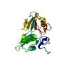

 PDBj
PDBj
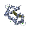
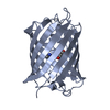
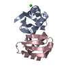
 Assembly
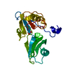


 Mass: 62.068 Da / Num. of mol.: 1 / Source method: obtained synthetically / Formula: C2H6O2
 Mass: 18.015 Da / Num. of mol.: 42 / Source method: isolated from a natural source / Formula: H2O
 Sample preparation
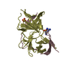
 / Beamline: ID23-2 / Wavelength: 0.97625 Å
 Processing