 Movie
Movie Controller

[English] 日本語
 Yorodumi
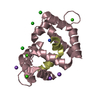
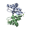
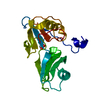
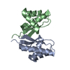
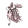
- PDB-2jzi: Structure of Calmodulin complexed with the Calmodulin Binding Dom... -
+ Open data

- Basic information
| Entry | Database: PDB / ID: 2jzi | ||||||
|---|---|---|---|---|---|---|---|
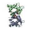
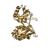
| Title | Structure of Calmodulin complexed with the Calmodulin Binding Domain of Calcineurin | ||||||
 Components Components |
| ||||||
 Keywords Keywords | METAL BINDING PROTEIN / Calcium binding protein / Acetylation / Methylation / Phosphoprotein / Ubl conjugation / Alternative splicing / Calmodulin-binding / Hydrolase / Iron / Metal-binding / Nucleus / Protein phosphatase / Zinc | ||||||
| Function / homology |  Function and homology information Function and homology informationnegative regulation of angiotensin-activated signaling pathway / regulation of cell proliferation involved in kidney morphogenesis / positive regulation of glomerulus development / negative regulation of calcium ion import across plasma membrane / negative regulation of signaling / positive regulation of saliva secretion / calmodulin-dependent protein phosphatase activity / calcineurin complex / positive regulation of connective tissue replacement / positive regulation of calcium ion-dependent exocytosis of neurotransmitter ...negative regulation of angiotensin-activated signaling pathway / regulation of cell proliferation involved in kidney morphogenesis / positive regulation of glomerulus development / negative regulation of calcium ion import across plasma membrane / negative regulation of signaling / positive regulation of saliva secretion / calmodulin-dependent protein phosphatase activity / calcineurin complex / positive regulation of connective tissue replacement / positive regulation of calcium ion-dependent exocytosis of neurotransmitter / positive regulation of calcium ion import across plasma membrane / positive regulation of cardiac muscle hypertrophy in response to stress / renal filtration / : / : / : / : / positive regulation of protein autophosphorylation / : / calcineurin-NFAT signaling cascade / positive regulation of calcineurin-NFAT signaling cascade / negative regulation of peptidyl-threonine phosphorylation / skeletal muscle tissue regeneration / : / type 3 metabotropic glutamate receptor binding / positive regulation of osteoclast differentiation / dephosphorylation / positive regulation of activated T cell proliferation / positive regulation of DNA binding / CaM pathway / protein dephosphorylation / extrinsic component of plasma membrane / Cam-PDE 1 activation / positive regulation of peptidyl-threonine phosphorylation / Sodium/Calcium exchangers / Calmodulin induced events / Reduction of cytosolic Ca++ levels / Activation of Ca-permeable Kainate Receptor / CREB1 phosphorylation through the activation of CaMKII/CaMKK/CaMKIV cascasde / Loss of phosphorylation of MECP2 at T308 / CREB1 phosphorylation through the activation of Adenylate Cyclase / negative regulation of high voltage-gated calcium channel activity / PKA activation / CaMK IV-mediated phosphorylation of CREB / protein-serine/threonine phosphatase / Glycogen breakdown (glycogenolysis) / response to corticosterone / negative regulation of ryanodine-sensitive calcium-release channel activity / Activation of RAC1 downstream of NMDARs / organelle localization by membrane tethering / CLEC7A (Dectin-1) induces NFAT activation / : / autophagosome membrane docking / regulation of synaptic vesicle exocytosis / negative regulation of calcium ion export across plasma membrane / regulation of cardiac muscle cell action potential / presynaptic endocytosis / Synthesis of IP3 and IP4 in the cytosol / positive regulation of protein serine/threonine kinase activity / epidermis development / Phase 0 - rapid depolarisation / Negative regulation of NMDA receptor-mediated neuronal transmission / protein serine/threonine phosphatase activity / Unblocking of NMDA receptors, glutamate binding and activation / calcineurin-mediated signaling / RHO GTPases activate PAKs / nitric-oxide synthase binding / regulation of cell communication by electrical coupling involved in cardiac conduction / Ion transport by P-type ATPases / adenylate cyclase binding / Uptake and function of anthrax toxins / protein phosphatase activator activity / positive regulation of osteoblast differentiation / regulation of ryanodine-sensitive calcium-release channel activity / Long-term potentiation / Calcineurin activates NFAT / keratinocyte differentiation / Regulation of MECP2 expression and activity / DARPP-32 events / Smooth Muscle Contraction / regulation of synaptic vesicle endocytosis / positive regulation of endocytosis / detection of calcium ion / catalytic complex / regulation of cardiac muscle contraction / postsynaptic modulation of chemical synaptic transmission / cellular response to interferon-beta / RHO GTPases activate IQGAPs / positive regulation of nitric-oxide synthase activity / phosphatidylinositol 3-kinase binding / activation of adenylate cyclase activity / calcium channel inhibitor activity / presynaptic cytosol / skeletal muscle fiber development / Activation of AMPK downstream of NMDARs / regulation of release of sequestered calcium ion into cytosol by sarcoplasmic reticulum / enzyme regulator activity / eNOS activation / Ion homeostasis / Tetrahydrobiopterin (BH4) synthesis, recycling, salvage and regulation Similarity search - Function | ||||||
| Biological species |  Homo sapiens (human) Homo sapiens (human) | ||||||
| Method | SOLUTION NMR / distance geometry | ||||||
 Authors Authors | Chyan, C. / Huang, J. / Irene, D. / Lin, T. | ||||||
 Citation Citation | Journal: To be Published Title: Structure of Calmodulin complexed with the Calmodulin Binding Domain of Calcineurin Authors: Chyan, C. / Huang, J. / Irene, D. / Lin, T. | ||||||
| History |
|
- Structure visualization
| Structure viewer | Molecule: MolmilJmol/JSmol |
|---|
- Downloads & links
-Download
| PDBx/mmCIF format | 2jzi.cif.gz | 1 MB | Display | PDBx/mmCIF format |
|---|---|---|---|---|
| PDB format | pdb2jzi.ent.gz | 877.6 KB | Display | PDB format |
| PDBx/mmJSON format | 2jzi.json.gz | Tree view | PDBx/mmJSON format | |
| Others |  Other downloads Other downloads |
-Validation report
| Arichive directory | https://data.pdbj.org/pub/pdb/validation_reports/jz/2jziftp://data.pdbj.org/pub/pdb/validation_reports/jz/2jzi | HTTPS FTP |
|---|
-Related structure data
| Similar structure data |
|---|


-Links
 PDBj
PDBj


- Assembly
| Deposited unit | 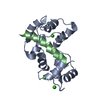
| |||||||||
|---|---|---|---|---|---|---|---|---|---|---|
| 1 |
| |||||||||
| NMR ensembles |
|
-Components
| #1: Protein | Mass: 16721.350 Da / Num. of mol.: 1 Source method: isolated from a genetically manipulated source Source: (gene. exp.) Homo sapiens (human) / Gene: CALM1, CALM, CAM, CAM1 / Production host:  |
|---|---|
| #2: Protein/peptide | Mass: 2820.496 Da / Num. of mol.: 1 Fragment: UNP residues 391-414, Calmodulin_binding_domain_of_Calcineurin Source method: isolated from a genetically manipulated source Source: (gene. exp.) Homo sapiens (human) / Gene: PPP3CA, CALNA, CNA / Production host: References: UniProt: Q08209, protein-serine/threonine phosphatase |
| #3: Chemical | ChemComp-CA /   Mass: 40.078 Da / Num. of mol.: 4 / Source method: obtained synthetically / Formula: Ca Mass: 40.078 Da / Num. of mol.: 4 / Source method: obtained synthetically / Formula: Ca |
-Experimental details
-Experiment
| Experiment | Method: SOLUTION NMR | ||||||||||||||||||||||||||||||||||||||||||||||||||||||||||||||||||||
|---|---|---|---|---|---|---|---|---|---|---|---|---|---|---|---|---|---|---|---|---|---|---|---|---|---|---|---|---|---|---|---|---|---|---|---|---|---|---|---|---|---|---|---|---|---|---|---|---|---|---|---|---|---|---|---|---|---|---|---|---|---|---|---|---|---|---|---|---|---|
| NMR experiment |
|
 HSQC
HSQC- Sample preparation
| Details | Contents: 1 mM [U-99% 13C; U-99% 15N] Calmodulin, 1 mM [U-99% 13C; U-99% 15N] Calmodulin binding domain of Calcineurin, 90% H2O/10% D2O Solvent system: 90% H2O/10% D2O | ||||||||||||
|---|---|---|---|---|---|---|---|---|---|---|---|---|---|
| Sample |
| ||||||||||||
| Sample conditions | pH: 6.5 / Temperature: 310 K |
-NMR measurement
| NMR spectrometer |
|
|---|
- Processing
| NMR software |
| ||||||||||||
|---|---|---|---|---|---|---|---|---|---|---|---|---|---|
| Refinement | Method: distance geometry / Software ordinal: 1 | ||||||||||||
| NMR constraints | Protein chi angle constraints total count: 0 / Protein other angle constraints total count: 284 / Protein phi angle constraints total count: 142 / Protein psi angle constraints total count: 142 | ||||||||||||
| NMR representative | Selection criteria: lowest energy | ||||||||||||
| NMR ensemble | Average torsion angle constraint violation: 0 ° Conformer selection criteria: back calculated data agree with experimental NOESY spectrum Conformers calculated total number: 100 / Conformers submitted total number: 20 / Maximum torsion angle constraint violation: 0 ° / Representative conformer: 1 / Torsion angle constraint violation method: TALOS |
