 Movie
Movie Controller

[English] 日本語
 Yorodumi
- PDB-5dow: Solution of the Variably-Twinned Structure of a Novel Calmodulin-... -
+ Open data

- Basic information
| Entry | Database: PDB / ID: 5dow | ||||||
|---|---|---|---|---|---|---|---|
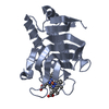
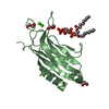
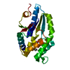
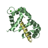
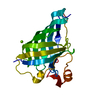
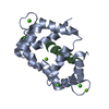
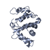
| Title | Solution of the Variably-Twinned Structure of a Novel Calmodulin-Peptide Complex in a Novel Configuration | ||||||
 Components Components |
| ||||||
 Keywords Keywords | CALCIUM BINDING PROTEIN / Calmodulin-Peptide Complex | ||||||
| Function / homology |  Function and homology information Function and homology informationInorganic anion exchange by SLC26 transporters / secondary active sulfate transmembrane transporter activity / intracellular pH elevation / : / : / : / : / positive regulation of protein autophosphorylation / : / negative regulation of peptidyl-threonine phosphorylation ...Inorganic anion exchange by SLC26 transporters / secondary active sulfate transmembrane transporter activity / intracellular pH elevation / : / : / : / : / positive regulation of protein autophosphorylation / : / negative regulation of peptidyl-threonine phosphorylation / chloride:bicarbonate antiporter activity / : / bicarbonate transmembrane transporter activity / type 3 metabotropic glutamate receptor binding / membrane hyperpolarization / chloride transmembrane transporter activity / positive regulation of DNA binding / CaM pathway / Cam-PDE 1 activation / positive regulation of peptidyl-threonine phosphorylation / Sodium/Calcium exchangers / Calmodulin induced events / Reduction of cytosolic Ca++ levels / Activation of Ca-permeable Kainate Receptor / CREB1 phosphorylation through the activation of CaMKII/CaMKK/CaMKIV cascasde / Loss of phosphorylation of MECP2 at T308 / CREB1 phosphorylation through the activation of Adenylate Cyclase / negative regulation of high voltage-gated calcium channel activity / PKA activation / CaMK IV-mediated phosphorylation of CREB / sperm capacitation / Glycogen breakdown (glycogenolysis) / response to corticosterone / negative regulation of ryanodine-sensitive calcium-release channel activity / Activation of RAC1 downstream of NMDARs / organelle localization by membrane tethering / CLEC7A (Dectin-1) induces NFAT activation / : / autophagosome membrane docking / regulation of synaptic vesicle exocytosis / negative regulation of calcium ion export across plasma membrane / regulation of cardiac muscle cell action potential / presynaptic endocytosis / Synthesis of IP3 and IP4 in the cytosol / positive regulation of protein serine/threonine kinase activity / Phase 0 - rapid depolarisation / Negative regulation of NMDA receptor-mediated neuronal transmission / Unblocking of NMDA receptors, glutamate binding and activation / calcineurin-mediated signaling / RHO GTPases activate PAKs / nitric-oxide synthase binding / regulation of cell communication by electrical coupling involved in cardiac conduction / Ion transport by P-type ATPases / adenylate cyclase binding / Uptake and function of anthrax toxins / protein phosphatase activator activity / regulation of ryanodine-sensitive calcium-release channel activity / Long-term potentiation / Calcineurin activates NFAT / Regulation of MECP2 expression and activity / DARPP-32 events / Smooth Muscle Contraction / regulation of synaptic vesicle endocytosis / detection of calcium ion / catalytic complex / regulation of cardiac muscle contraction / cellular response to interferon-beta / RHO GTPases activate IQGAPs / positive regulation of nitric-oxide synthase activity / phosphatidylinositol 3-kinase binding / activation of adenylate cyclase activity / calcium channel inhibitor activity / presynaptic cytosol / Activation of AMPK downstream of NMDARs / regulation of release of sequestered calcium ion into cytosol by sarcoplasmic reticulum / enzyme regulator activity / eNOS activation / Ion homeostasis / Tetrahydrobiopterin (BH4) synthesis, recycling, salvage and regulation / regulation of calcium-mediated signaling / Protein methylation / titin binding / regulation of cardiac muscle contraction by regulation of the release of sequestered calcium ion / voltage-gated potassium channel complex / FCERI mediated Ca+2 mobilization / calcium channel complex / substantia nigra development / regulation of heart rate / FCGR3A-mediated IL10 synthesis / calyx of Held / cellular response to cAMP / Ras activation upon Ca2+ influx through NMDA receptor / Antigen activates B Cell Receptor (BCR) leading to generation of second messengers / response to amphetamine / nitric-oxide synthase regulator activity / adenylate cyclase activator activity / protein serine/threonine kinase activator activity / VEGFR2 mediated cell proliferation / VEGFR2 mediated vascular permeability / regulation of cytokinesis Similarity search - Function | ||||||
| Biological species |  Homo sapiens (human) Homo sapiens (human) | ||||||
| Method |  X-RAY DIFFRACTION / MOLECULAR REPLACEMENT / molecular replacement / Resolution: 1.7 Å X-RAY DIFFRACTION / MOLECULAR REPLACEMENT / molecular replacement / Resolution: 1.7 Å | ||||||
 Authors Authors | Keller, J.P. | ||||||
 Citation Citation | Journal: Acta Crystallogr D Struct Biol / Year: 2017 Title: Solution of the structure of a calmodulin-peptide complex in a novel configuration from a variably twinned data set. Authors: Keller, J.P. | ||||||
| History |
|
- Structure visualization
| Structure viewer | Molecule: MolmilJmol/JSmol |
|---|
- Downloads & links
-Download
| PDBx/mmCIF format | 5dow.cif.gz | 350.3 KB | Display | PDBx/mmCIF format |
|---|---|---|---|---|
| PDB format | pdb5dow.ent.gz | 286.3 KB | Display | PDB format |
| PDBx/mmJSON format | 5dow.json.gz | Tree view | PDBx/mmJSON format | |
| Others |  Other downloads Other downloads |
-Validation report
| Arichive directory | https://data.pdbj.org/pub/pdb/validation_reports/do/5dowftp://data.pdbj.org/pub/pdb/validation_reports/do/5dow | HTTPS FTP |
|---|
-Related structure data
| Similar structure data |
|---|


-Links
 PDBj
PDBj


- Assembly
| Deposited unit | 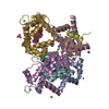
| |||||||||
|---|---|---|---|---|---|---|---|---|---|---|
| 1 | 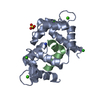
| |||||||||
| 2 | 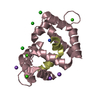
| |||||||||
| 3 | 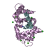
| |||||||||
| 4 | 
| |||||||||
| Unit cell |
| |||||||||
| Components on special symmetry positions |
|
-Components
-Protein / Protein/peptide , 2 types, 8 molecules ACEGBDFH
| #1: Protein | Mass: 16769.436 Da / Num. of mol.: 4 Source method: isolated from a genetically manipulated source Source: (gene. exp.) Homo sapiens (human)Gene: CALM1, CALM, CAM, CAM1, CALM2, CAM2, CAMB, CALM3, CALML2, CAM3, CAMC, CAMIII Plasmid: pET24 / Production host:  #2: Protein/peptide | Mass: 2559.262 Da / Num. of mol.: 4 / Fragment: peptide (UNP residues 563-583) / Source method: obtained synthetically / Source: (synth.) |
|---|
-Non-polymers , 5 types, 1128 molecules 


| #3: Chemical |  Mass: 96.063 Da / Num. of mol.: 2 / Source method: obtained synthetically / Formula: SO4 Mass: 96.063 Da / Num. of mol.: 2 / Source method: obtained synthetically / Formula: SO4#4: Chemical | ChemComp-CA /  Mass: 40.078 Da / Num. of mol.: 16 / Source method: obtained synthetically / Formula: Ca Mass: 40.078 Da / Num. of mol.: 16 / Source method: obtained synthetically / Formula: Ca#5: Chemical | ChemComp-CL /  Mass: 35.453 Da / Num. of mol.: 11 / Source method: obtained synthetically / Formula: Cl Mass: 35.453 Da / Num. of mol.: 11 / Source method: obtained synthetically / Formula: Cl#6: Chemical | ChemComp-NA /  Mass: 22.990 Da / Num. of mol.: 5 / Source method: obtained synthetically / Formula: Na Mass: 22.990 Da / Num. of mol.: 5 / Source method: obtained synthetically / Formula: Na#7: Water | ChemComp-HOH / | Mass: 18.015 Da / Num. of mol.: 1094 / Source method: isolated from a natural source / Formula: H2O |
|---|
-Details
| Has protein modification | Y |
|---|
-Experimental details
-Experiment
| Experiment | Method: X-RAY DIFFRACTION / Number of used crystals: 1 |
|---|
- Sample preparation
| Crystal | Density Matthews: 2.36 Å3/Da / Density % sol: 47.93 % |
|---|---|
| Crystal grow | Temperature: 298 K / Method: vapor diffusion, hanging drop / pH: 7.5 / Details: ammonium sulfate, lithium sulfate, Tris/HEPES / PH range: 7.0 - 8.0 |
-Data collection
| Diffraction | Mean temperature: 100 K | ||||||||||||||||||
|---|---|---|---|---|---|---|---|---|---|---|---|---|---|---|---|---|---|---|---|
| Diffraction source | Source: ROTATING ANODE / Type: RIGAKU MICROMAX-007 HF / Wavelength: 1.5418 Å | ||||||||||||||||||
| Detector | Type: RIGAKU SATURN 944 / Detector: CCD / Date: Jan 27, 2015 / Details: Varimax CuHF | ||||||||||||||||||
| Radiation | Protocol: SINGLE WAVELENGTH / Monochromatic (M) / Laue (L): M / Scattering type: x-ray | ||||||||||||||||||
| Radiation wavelength | Wavelength: 1.5418 Å / Relative weight: 1 | ||||||||||||||||||
| Reflection twin |
| ||||||||||||||||||
| Reflection | Resolution: 1.7→22.81 Å / Num. obs: 76360 / % possible obs: 96.5 % / Redundancy: 21.5 % / CC1/2: 0.998 / Rmerge(I) obs: 0.107 / Rpim(I) all: 0.021 / Net I/σ(I): 22.1 / Num. measured all: 1645262 / Scaling rejects: 2402 | ||||||||||||||||||
| Reflection shell |
|
-Phasing
| Phasing | Method: molecular replacement | |||||||||
|---|---|---|---|---|---|---|---|---|---|---|
| Phasing MR | Model details: Phaser MODE: MR_AUTO
|
- Processing
| Software |
| ||||||||||||||||||||||||||||||||||||||||||||||||||||||||||||||||||||||||||||||||||||||||||||||||||||||||||||||||||||||||||||||||||||||||||||||||||||||||||||||||||||||||||||||||||||||
|---|---|---|---|---|---|---|---|---|---|---|---|---|---|---|---|---|---|---|---|---|---|---|---|---|---|---|---|---|---|---|---|---|---|---|---|---|---|---|---|---|---|---|---|---|---|---|---|---|---|---|---|---|---|---|---|---|---|---|---|---|---|---|---|---|---|---|---|---|---|---|---|---|---|---|---|---|---|---|---|---|---|---|---|---|---|---|---|---|---|---|---|---|---|---|---|---|---|---|---|---|---|---|---|---|---|---|---|---|---|---|---|---|---|---|---|---|---|---|---|---|---|---|---|---|---|---|---|---|---|---|---|---|---|---|---|---|---|---|---|---|---|---|---|---|---|---|---|---|---|---|---|---|---|---|---|---|---|---|---|---|---|---|---|---|---|---|---|---|---|---|---|---|---|---|---|---|---|---|---|---|---|---|---|
| Refinement | Method to determine structure: MOLECULAR REPLACEMENT / Resolution: 1.7→22.81 Å / Cor.coef. Fo:Fc: 0.982 / Cor.coef. Fo:Fc free: 0.97 / SU B: 2.932 / SU ML: 0.046 / Cross valid method: THROUGHOUT / ESU R: 0.029 / ESU R Free: 0.016 / Stereochemistry target values: MAXIMUM LIKELIHOOD / Details: HYDROGENS HAVE BEEN ADDED IN THE RIDING POSITIONS
| ||||||||||||||||||||||||||||||||||||||||||||||||||||||||||||||||||||||||||||||||||||||||||||||||||||||||||||||||||||||||||||||||||||||||||||||||||||||||||||||||||||||||||||||||||||||
| Solvent computation | Ion probe radii: 0.8 Å / Shrinkage radii: 0.8 Å / VDW probe radii: 1.2 Å / Solvent model: MASK | ||||||||||||||||||||||||||||||||||||||||||||||||||||||||||||||||||||||||||||||||||||||||||||||||||||||||||||||||||||||||||||||||||||||||||||||||||||||||||||||||||||||||||||||||||||||
| Displacement parameters | Biso mean: 21.167 Å2
| ||||||||||||||||||||||||||||||||||||||||||||||||||||||||||||||||||||||||||||||||||||||||||||||||||||||||||||||||||||||||||||||||||||||||||||||||||||||||||||||||||||||||||||||||||||||
| Refinement step | Cycle: LAST / Resolution: 1.7→22.81 Å
| ||||||||||||||||||||||||||||||||||||||||||||||||||||||||||||||||||||||||||||||||||||||||||||||||||||||||||||||||||||||||||||||||||||||||||||||||||||||||||||||||||||||||||||||||||||||
| Refine LS restraints |
|