 Movie
Movie Controller


+ Open data

- Basic information
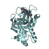
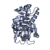
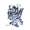
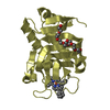
| Entry | Database: PDB / ID: 3ell | ||||||
|---|---|---|---|---|---|---|---|
| Title | Structure of the hemophore from Pseudomonas aeruginosa (HasAp) | ||||||
 Components Components | HasAp (Heme acquisition protein HasAp) | ||||||
 Keywords Keywords | HEME BINDING PROTEIN / alpha beta protein / heme-binding protein | ||||||
| Function / homology |  Function and homology information Function and homology information | ||||||
| Biological species |   Pseudomonas aeruginosa (bacteria) Pseudomonas aeruginosa (bacteria) | ||||||
| Method |  X-RAY DIFFRACTION / MOLECULAR REPLACEMENT / Resolution: 1.7 Å X-RAY DIFFRACTION / MOLECULAR REPLACEMENT / Resolution: 1.7 Å | ||||||
 Authors Authors | Schonbrunn, E. / Rivera, M. | ||||||
 Citation Citation | Journal: Biochemistry / Year: 2009 Title: Structural characterization of the hemophore HasAp from Pseudomonas aeruginosa: NMR spectroscopy reveals protein-protein interactions between Holo-HasAp and hemoglobin. Authors: Alontaga, A.Y. / Rodriguez, J.C. / Schonbrunn, E. / Becker, A. / Funke, T. / Yukl, E.T. / Hayashi, T. / Stobaugh, J. / Moenne-Loccoz, P. / Rivera, M. | ||||||
| History |
|
- Structure visualization
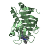
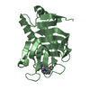
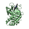
| Structure viewer | Molecule: MolmilJmol/JSmol |
|---|
- Downloads & links
-Download
| PDBx/mmCIF format | 3ell.cif.gz | 87 KB | Display | PDBx/mmCIF format |
|---|---|---|---|---|
| PDB format | pdb3ell.ent.gz | 65 KB | Display | PDB format |
| PDBx/mmJSON format | 3ell.json.gz | Tree view | PDBx/mmJSON format | |
| Others |  Other downloads Other downloads |
-Validation report
| Arichive directory | https://data.pdbj.org/pub/pdb/validation_reports/el/3ellftp://data.pdbj.org/pub/pdb/validation_reports/el/3ell | HTTPS FTP |
|---|
-Related structure data
| Related structure data | 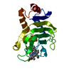 1b2vS S: Starting model for refinement |
|---|---|
| Similar structure data |
-Links
 PDBj
PDBj- Assembly
| Deposited unit | 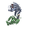
| ||||||||
|---|---|---|---|---|---|---|---|---|---|
| 1 | 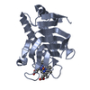
| ||||||||
| 2 | 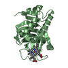
| ||||||||
| Unit cell |
|
-Components
| #1: Protein | Mass: 18901.535 Da / Num. of mol.: 2 / Fragment: HasAp (UNP residues 1 - 184) Source method: isolated from a genetically manipulated source Source: (gene. exp.) Pseudomonas aeruginosa (bacteria) / Gene: hasAp, PA3407 / Plasmid: pET11a / Production host: #2: Chemical |   Mass: 616.487 Da / Num. of mol.: 2 / Source method: obtained synthetically / Formula: C34H32FeN4O4 Mass: 616.487 Da / Num. of mol.: 2 / Source method: obtained synthetically / Formula: C34H32FeN4O4#3: Water | ChemComp-HOH / |  Mass: 18.015 Da / Num. of mol.: 250 / Source method: isolated from a natural source / Formula: H2O Mass: 18.015 Da / Num. of mol.: 250 / Source method: isolated from a natural source / Formula: H2O |
|---|
-Experimental details
-Experiment
| Experiment | Method: X-RAY DIFFRACTION / Number of used crystals: 1 |
|---|
- Sample preparation
| Crystal | Density Matthews: 2.45 Å3/Da / Density % sol: 49.9 % |
|---|---|
| Crystal grow | Temperature: 296 K / Method: vapor diffusion, hanging drop / pH: 6 Details: 100 mM MES buffer (pH 6.0), 100 mM NH4Cl, and 80% PEG 400, VAPOR DIFFUSION, HANGING DROP, temperature 296K |
-Data collection
| Diffraction | Mean temperature: 93 K |
|---|---|
| Diffraction source | Source: ROTATING ANODE / Type: RIGAKU MICROMAX-007 HF / Wavelength: 1.5418 Å |
| Detector | Type: RIGAKU RAXIS HTC / Detector: IMAGE PLATE / Date: Oct 1, 2007 / Details: mirrors |
| Radiation | Monochromator: mirrors / Protocol: SINGLE WAVELENGTH / Monochromatic (M) / Laue (L): M / Scattering type: x-ray |
| Radiation wavelength | Wavelength: 1.5418 Å / Relative weight: 1 |
| Reflection | Resolution: 1.7→20 Å / Num. all: 36833 / Num. obs: 36833 / % possible obs: 93.1 % / Observed criterion σ(F): 0 / Observed criterion σ(I): -3 / Redundancy: 2.2 % / Rmerge(I) obs: 0.031 / Net I/σ(I): 22.2 |
| Reflection shell | Resolution: 1.7→1.8 Å / Redundancy: 1.9 % / Rmerge(I) obs: 0.063 / Mean I/σ(I) obs: 12.7 / Num. unique all: 5430 / % possible all: 87.6 |
- Processing
| Software |
| |||||||||||||||||||||||||
|---|---|---|---|---|---|---|---|---|---|---|---|---|---|---|---|---|---|---|---|---|---|---|---|---|---|---|
| Refinement | Method to determine structure: MOLECULAR REPLACEMENT Starting model: PDB entry 1B2V Resolution: 1.7→20 Å / Cross valid method: THROUGHOUT / σ(F): 0 / σ(I): -3 / Stereochemistry target values: Engh & Huber
| |||||||||||||||||||||||||
| Refine analyze |
| |||||||||||||||||||||||||
| Refinement step | Cycle: LAST / Resolution: 1.7→20 Å
| |||||||||||||||||||||||||
| Refine LS restraints |
|
