 Movie
Movie Controller


+ Open data

- Basic information
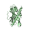
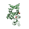
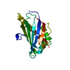
| Entry | Database: PDB / ID: 5xic | ||||||
|---|---|---|---|---|---|---|---|
| Title | Crystal Structure of HasAp with Fe-5,10,15-triphenylporphyrin | ||||||
 Components Components | Heme acquisition protein HasAp | ||||||
 Keywords Keywords | TRANSPORT PROTEIN / HEME ACQUISITION PROTEIN | ||||||
| Function / homology |  Function and homology information Function and homology information | ||||||
| Biological species |  Pseudomonas aeruginosa str. PAO1 (bacteria) Pseudomonas aeruginosa str. PAO1 (bacteria) | ||||||
| Method |  X-RAY DIFFRACTION / SYNCHROTRON / MOLECULAR REPLACEMENT / Resolution: 1.45 Å X-RAY DIFFRACTION / SYNCHROTRON / MOLECULAR REPLACEMENT / Resolution: 1.45 Å | ||||||
 Authors Authors | Shoji, O. / Uehara, H. / Sugimoto, H. / Shiro, Y. / Watanabe, Y. | ||||||
 Citation Citation | Journal: Angew. Chem. Int. Ed. Engl. / Year: 2017 Title: Structures of the Heme Acquisition Protein HasA with Iron(III)-5,15-Diphenylporphyrin and Derivatives Thereof as an Artificial Prosthetic Group Authors: Uehara, H. / Shisaka, Y. / Nishimura, T. / Sugimoto, H. / Shiro, Y. / Miyake, Y. / Shinokubo, H. / Watanabe, Y. / Shoji, O. | ||||||
| History |
|
- Structure visualization
| Structure viewer | Molecule: MolmilJmol/JSmol |
|---|
- Downloads & links
-Download
| PDBx/mmCIF format | 5xic.cif.gz | 165.8 KB | Display | PDBx/mmCIF format |
|---|---|---|---|---|
| PDB format | pdb5xic.ent.gz | 132.1 KB | Display | PDB format |
| PDBx/mmJSON format | 5xic.json.gz | Tree view | PDBx/mmJSON format | |
| Others |  Other downloads Other downloads |
-Validation report
| Arichive directory | https://data.pdbj.org/pub/pdb/validation_reports/xi/5xicftp://data.pdbj.org/pub/pdb/validation_reports/xi/5xic | HTTPS FTP |
|---|
-Related structure data
| Related structure data |  5xa4C  5xibC  5xieC  5xkbC  3w8oS S: Starting model for refinement C: citing same article ( |
|---|---|
| Similar structure data |


-Links
 PDBj
PDBj- Assembly
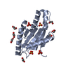
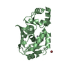
| Deposited unit | 
| ||||||||
|---|---|---|---|---|---|---|---|---|---|
| 1 | 
| ||||||||
| 2 | 
| ||||||||
| 3 | 
| ||||||||
| 4 | 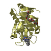
| ||||||||
| Unit cell |
|
-Components
| #1: Protein | Mass: 18901.535 Da / Num. of mol.: 4 / Fragment: UNP Residues 1-184 / Mutation: Wild-type Source method: isolated from a genetically manipulated source Source: (gene. exp.) Pseudomonas aeruginosa str. PAO1 (bacteria)Strain: PAO1 / Gene: hasAp, PA3407 / Plasmid: PQE30 / Production host: #2: Chemical | ChemComp-WXP / 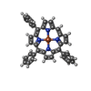  Mass: 592.469 Da / Num. of mol.: 4 / Source method: obtained synthetically / Formula: C38H24FeN4 Mass: 592.469 Da / Num. of mol.: 4 / Source method: obtained synthetically / Formula: C38H24FeN4#3: Chemical | ChemComp-PE8 /   Mass: 370.436 Da / Num. of mol.: 4 / Source method: obtained synthetically / Formula: C16H34O9 Mass: 370.436 Da / Num. of mol.: 4 / Source method: obtained synthetically / Formula: C16H34O9#4: Water | ChemComp-HOH / |  Mass: 18.015 Da / Num. of mol.: 476 / Source method: isolated from a natural source / Formula: H2O Mass: 18.015 Da / Num. of mol.: 476 / Source method: isolated from a natural source / Formula: H2O |
|---|
-Experimental details
-Experiment
| Experiment | Method: X-RAY DIFFRACTION / Number of used crystals: 1 |
|---|
- Sample preparation
| Crystal | Density Matthews: 2.1 Å3/Da / Density % sol: 41.53 % / Mosaicity: 0.831 ° |
|---|---|
| Crystal grow | Temperature: 293 K / Method: vapor diffusion, sitting drop / pH: 8.5 Details: 15% v/v PEG 400, 50mM Tris-HCl (pH8.5), 50mM KPi buffer (pH7.0), 100mM MgCl2, 15% v/v PEG 400 |
-Data collection
| Diffraction | Mean temperature: 100 K | |||||||||||||||||||||||||||||||||||||||||||||||||||||||||||||||||||||||||||||||||||||||||||||||||||
|---|---|---|---|---|---|---|---|---|---|---|---|---|---|---|---|---|---|---|---|---|---|---|---|---|---|---|---|---|---|---|---|---|---|---|---|---|---|---|---|---|---|---|---|---|---|---|---|---|---|---|---|---|---|---|---|---|---|---|---|---|---|---|---|---|---|---|---|---|---|---|---|---|---|---|---|---|---|---|---|---|---|---|---|---|---|---|---|---|---|---|---|---|---|---|---|---|---|---|---|---|
| Diffraction source | Source: SYNCHROTRON / Site: SPring-8  / Beamline: BL26B2 / Wavelength: 1 Å / Beamline: BL26B2 / Wavelength: 1 Å | |||||||||||||||||||||||||||||||||||||||||||||||||||||||||||||||||||||||||||||||||||||||||||||||||||
| Detector | Type: MARMOSAIC 225 mm CCD / Detector: CCD / Date: Nov 9, 2016 | |||||||||||||||||||||||||||||||||||||||||||||||||||||||||||||||||||||||||||||||||||||||||||||||||||
| Radiation | Monochromator: Si 111 CHANNEL / Protocol: SINGLE WAVELENGTH / Monochromatic (M) / Laue (L): M / Scattering type: x-ray | |||||||||||||||||||||||||||||||||||||||||||||||||||||||||||||||||||||||||||||||||||||||||||||||||||
| Radiation wavelength | Wavelength: 1 Å / Relative weight: 1 | |||||||||||||||||||||||||||||||||||||||||||||||||||||||||||||||||||||||||||||||||||||||||||||||||||
| Reflection | Resolution: 1.45→50 Å / Num. obs: 107752 / % possible obs: 96.2 % / Redundancy: 3.1 % / Rmerge(I) obs: 0.028 / Rpim(I) all: 0.019 / Rrim(I) all: 0.034 / Χ2: 0.796 / Net I/σ(I): 16.6 / Num. measured all: 330905 | |||||||||||||||||||||||||||||||||||||||||||||||||||||||||||||||||||||||||||||||||||||||||||||||||||
| Reflection shell | Diffraction-ID: 1
|
- Processing
| Software |
| ||||||||||||||||||||||||||||||||||||||||||||||||||||||||||||
|---|---|---|---|---|---|---|---|---|---|---|---|---|---|---|---|---|---|---|---|---|---|---|---|---|---|---|---|---|---|---|---|---|---|---|---|---|---|---|---|---|---|---|---|---|---|---|---|---|---|---|---|---|---|---|---|---|---|---|---|---|---|
| Refinement | Method to determine structure: MOLECULAR REPLACEMENT Starting model: 3W8O Resolution: 1.45→19.48 Å / Cor.coef. Fo:Fc: 0.954 / Cor.coef. Fo:Fc free: 0.944 / WRfactor Rfree: 0.232 / WRfactor Rwork: 0.2028 / FOM work R set: 0.8598 / SU B: 1.36 / SU ML: 0.053 / SU R Cruickshank DPI: 0.0831 / SU Rfree: 0.0813 / Cross valid method: THROUGHOUT / σ(F): 0 / ESU R: 0.083 / ESU R Free: 0.081 / Stereochemistry target values: MAXIMUM LIKELIHOOD Details: HYDROGENS HAVE BEEN ADDED IN THE RIDING POSITIONS U VALUES : REFINED INDIVIDUALLY
| ||||||||||||||||||||||||||||||||||||||||||||||||||||||||||||
| Solvent computation | Ion probe radii: 0.8 Å / Shrinkage radii: 0.8 Å / VDW probe radii: 1.2 Å / Solvent model: MASK | ||||||||||||||||||||||||||||||||||||||||||||||||||||||||||||
| Displacement parameters | Biso max: 46.14 Å2 / Biso mean: 13.68 Å2 / Biso min: 6.67 Å2
| ||||||||||||||||||||||||||||||||||||||||||||||||||||||||||||
| Refinement step | Cycle: final / Resolution: 1.45→19.48 Å
| ||||||||||||||||||||||||||||||||||||||||||||||||||||||||||||
| Refine LS restraints |
| ||||||||||||||||||||||||||||||||||||||||||||||||||||||||||||
| LS refinement shell | Resolution: 1.45→1.487 Å / Rfactor Rfree error: 0 / Total num. of bins used: 20
|
