 Movie
Movie Controller

[English] 日本語
 Yorodumi
- PDB-3eop: Crystal Structure of the DUF55 domain of human thymocyte nuclear ... -
+ Open data

- Basic information
| Entry | Database: PDB / ID: 3eop | ||||||
|---|---|---|---|---|---|---|---|
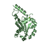
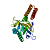
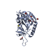
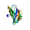
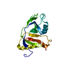
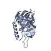
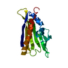
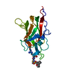
| Title | Crystal Structure of the DUF55 domain of human thymocyte nuclear protein 1 | ||||||
 Components Components | Thymocyte nuclear protein 1 | ||||||
 Keywords Keywords | UNKNOWN FUNCTION / Nucleus / Phosphoprotein | ||||||
| Function / homology |  Function and homology information Function and homology information | ||||||
| Biological species |  Homo sapiens (human) Homo sapiens (human) | ||||||
| Method |  X-RAY DIFFRACTION / SYNCHROTRON / MOLECULAR REPLACEMENT / molecular replacement / Resolution: 2.3 Å X-RAY DIFFRACTION / SYNCHROTRON / MOLECULAR REPLACEMENT / molecular replacement / Resolution: 2.3 Å | ||||||
 Authors Authors | Yu, F. / Song, A. / Xu, C. / Sun, L. / Li, L. / Tang, L. / Hu, H. / He, J. | ||||||
 Citation Citation | Journal: Acta Crystallogr.,Sect.D / Year: 2009 Title: Determining the DUF55-domain structure of human thymocyte nuclear protein 1 from crystals partially twinned by tetartohedry Authors: Yu, F. / Song, A. / Xu, C. / Sun, L. / Li, J. / Tang, L. / Yu, M. / Yeates, T.O. / Hu, H. / He, J. | ||||||
| History |
|
- Structure visualization
| Structure viewer | Molecule: MolmilJmol/JSmol |
|---|
- Downloads & links
-Download
| PDBx/mmCIF format | 3eop.cif.gz | 80.8 KB | Display | PDBx/mmCIF format |
|---|---|---|---|---|
| PDB format | pdb3eop.ent.gz | 60.7 KB | Display | PDB format |
| PDBx/mmJSON format | 3eop.json.gz | Tree view | PDBx/mmJSON format | |
| Others |  Other downloads Other downloads |
-Validation report
| Arichive directory | https://data.pdbj.org/pub/pdb/validation_reports/eo/3eopftp://data.pdbj.org/pub/pdb/validation_reports/eo/3eop | HTTPS FTP |
|---|
-Related structure data
| Related structure data | 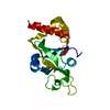 2ar1S S: Starting model for refinement |
|---|---|
| Similar structure data |


-Links
 PDBj
PDBj
- Assembly
| Deposited unit | 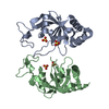
| ||||||||
|---|---|---|---|---|---|---|---|---|---|
| 1 | 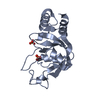
| ||||||||
| 2 | 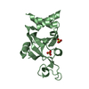
| ||||||||
| Unit cell |
|
-Components
| #1: Protein | Mass: 20770.738 Da / Num. of mol.: 2 / Fragment: DUF55 domain, UNP residues 55-221 Source method: isolated from a genetically manipulated source Source: (gene. exp.) Homo sapiens (human) / Plasmid: pET22 / Production host:  #2: Chemical | ChemComp-SO4 /   Mass: 96.063 Da / Num. of mol.: 4 / Source method: obtained synthetically / Formula: SO4 Mass: 96.063 Da / Num. of mol.: 4 / Source method: obtained synthetically / Formula: SO4#3: Water | ChemComp-HOH / |  Mass: 18.015 Da / Num. of mol.: 37 / Source method: isolated from a natural source / Formula: H2O Mass: 18.015 Da / Num. of mol.: 37 / Source method: isolated from a natural source / Formula: H2OHas protein modification | Y | |
|---|
-Experimental details
-Experiment
| Experiment | Method: X-RAY DIFFRACTION / Number of used crystals: 1 |
|---|
- Sample preparation
| Crystal | Density Matthews: 2.23 Å3/Da / Density % sol: 44.96 % |
|---|---|
| Crystal grow | Temperature: 291 K / Method: vapor diffusion, hanging drop / pH: 10.9 Details: 0.1M NaAc (pH 4.0-4.8), 28% PEG 2000 MME, 200mM (NH4)2SO4, 3% 1,6-diaminohexane as additive, pH 10.9, VAPOR DIFFUSION, HANGING DROP, temperature 291K |
-Data collection
| Diffraction | Mean temperature: 100 K |
|---|---|
| Diffraction source | Source: SYNCHROTRON / Site: BSRF  / Beamline: 3W1A / Wavelength: 1 Å / Beamline: 3W1A / Wavelength: 1 Å |
| Detector | Type: MAR CCD 165 mm / Detector: CCD / Date: Mar 7, 2008 |
| Radiation | Protocol: SINGLE WAVELENGTH / Monochromatic (M) / Laue (L): M / Scattering type: x-ray |
| Radiation wavelength | Wavelength: 1 Å / Relative weight: 1 |
| Reflection | Resolution: 2.3→44.41 Å / Num. all: 15997 / Num. obs: 15931 / % possible obs: 99.4 % / Redundancy: 4.8 % / Biso Wilson estimate: 40.06 Å2 / Rmerge(I) obs: 0.056 |
| Reflection shell | Resolution: 2.3→2.42 Å / Redundancy: 4.5 % / Rmerge(I) obs: 0.291 / Num. unique all: 2326 / % possible all: 98.8 |
-Phasing
| Phasing | Method: molecular replacement |
|---|
- Processing
| Software |
| ||||||||||||||||||||||||||||||||||||
|---|---|---|---|---|---|---|---|---|---|---|---|---|---|---|---|---|---|---|---|---|---|---|---|---|---|---|---|---|---|---|---|---|---|---|---|---|---|
| Refinement | Method to determine structure: MOLECULAR REPLACEMENT Starting model: PDB entry 2ar1 Resolution: 2.3→44.39 Å / Occupancy max: 1 / Occupancy min: 1 / Data cutoff high absF: 0 / σ(F): 0 Details: Thin resolution shells. Quadruplets of twin related reflections were kept together in either the test set or the refinement set throughout refinement.
| ||||||||||||||||||||||||||||||||||||
| Solvent computation | Bsol: 59.208 Å2 | ||||||||||||||||||||||||||||||||||||
| Displacement parameters | Biso max: 68.11 Å2 / Biso mean: 36.2394 Å2 / Biso min: 5.91 Å2
| ||||||||||||||||||||||||||||||||||||
| Refine analyze |
| ||||||||||||||||||||||||||||||||||||
| Refinement step | Cycle: LAST / Resolution: 2.3→44.39 Å
| ||||||||||||||||||||||||||||||||||||
| Refine LS restraints |
| ||||||||||||||||||||||||||||||||||||
| LS refinement shell | Resolution: 2.3→2.38 Å
| ||||||||||||||||||||||||||||||||||||
| Xplor file |
|
