 Movie
Movie Controller


+ Open data

- Basic information
| Entry | Database: PDB / ID: 5o1y | ||||||
|---|---|---|---|---|---|---|---|
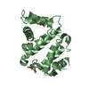
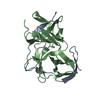
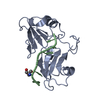
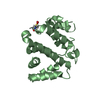
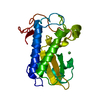
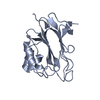
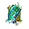
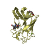
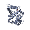
| Title | Structure of Nrd1 RNA binding domain in complex with RNA (GUAA) | ||||||
 Components Components |
| ||||||
 Keywords Keywords | TRANSCRIPTION / Nrd1 / Nrd1 complex / RRM / RNA-binding / non-coding transcription | ||||||
| Function / homology |  Function and homology information Function and homology informationtranscription regulatory region RNA binding / antisense RNA transcript catabolic process / Nrd1 complex / termination of RNA polymerase II transcription, exosome-dependent / sno(s)RNA 3'-end processing / CUT catabolic process / snRNA 3'-end processing / tRNA 3'-end processing / nuclear mRNA surveillance / mRNA 3'-end processing ...transcription regulatory region RNA binding / antisense RNA transcript catabolic process / Nrd1 complex / termination of RNA polymerase II transcription, exosome-dependent / sno(s)RNA 3'-end processing / CUT catabolic process / snRNA 3'-end processing / tRNA 3'-end processing / nuclear mRNA surveillance / mRNA 3'-end processing / protein domain specific binding / mRNA binding / RNA binding / nucleus Similarity search - Function | ||||||
| Biological species |   | ||||||
| Method |  X-RAY DIFFRACTION / SYNCHROTRON / MOLECULAR REPLACEMENT / Resolution: 2.45 Å X-RAY DIFFRACTION / SYNCHROTRON / MOLECULAR REPLACEMENT / Resolution: 2.45 Å | ||||||
 Authors Authors | Franco-Echevarria, E. / Perez-Canadillas, J.M. / Gonzalez, B. | ||||||
| Funding support |  Spain, 1items Spain, 1items
| ||||||
 Citation Citation | Journal: Nucleic Acids Res. / Year: 2017 Title: The structure of transcription termination factor Nrd1 reveals an original mode for GUAA recognition. Authors: Franco-Echevarria, E. / Gonzalez-Polo, N. / Zorrilla, S. / Martinez-Lumbreras, S. / Santiveri, C.M. / Campos-Olivas, R. / Sanchez, M. / Calvo, O. / Gonzalez, B. / Perez-Canadillas, J.M. | ||||||
| History |
|
- Structure visualization
| Structure viewer | Molecule: MolmilJmol/JSmol |
|---|
- Downloads & links
-Download
| PDBx/mmCIF format | 5o1y.cif.gz | 52.6 KB | Display | PDBx/mmCIF format |
|---|---|---|---|---|
| PDB format | pdb5o1y.ent.gz | 34.8 KB | Display | PDB format |
| PDBx/mmJSON format | 5o1y.json.gz | Tree view | PDBx/mmJSON format | |
| Others |  Other downloads Other downloads |
-Validation report
| Arichive directory | https://data.pdbj.org/pub/pdb/validation_reports/o1/5o1yftp://data.pdbj.org/pub/pdb/validation_reports/o1/5o1y | HTTPS FTP |
|---|
-Related structure data
| Related structure data | 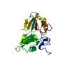 5o1tC 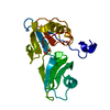 5o1wSC 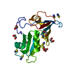 5o1xC  5o1zC  5o20C S: Starting model for refinement C: citing same article ( |
|---|---|
| Similar structure data |

-Links
 PDBj
PDBj
- Assembly
| Deposited unit | 
| ||||||||
|---|---|---|---|---|---|---|---|---|---|
| 1 |
| ||||||||
| Unit cell |
|
-Components
| #1: Protein | Mass: 20108.586 Da / Num. of mol.: 1 Source method: isolated from a genetically manipulated source Source: (gene. exp.) Gene: NRD1, YNL251C, N0868 / Production host:  | ||||
|---|---|---|---|---|---|
| #2: RNA chain | Mass: 1264.826 Da / Num. of mol.: 1 / Source method: obtained synthetically / Source: (synth.) | ||||
| #3: Chemical |   Mass: 62.068 Da / Num. of mol.: 2 / Source method: obtained synthetically / Formula: C2H6O2 Mass: 62.068 Da / Num. of mol.: 2 / Source method: obtained synthetically / Formula: C2H6O2#4: Chemical | ChemComp-TRS / |   Mass: 122.143 Da / Num. of mol.: 1 / Source method: obtained synthetically / Formula: C4H12NO3 / Comment: pH buffer*YM Mass: 122.143 Da / Num. of mol.: 1 / Source method: obtained synthetically / Formula: C4H12NO3 / Comment: pH buffer*YM#5: Water | ChemComp-HOH / |  Mass: 18.015 Da / Num. of mol.: 38 / Source method: isolated from a natural source / Formula: H2O Mass: 18.015 Da / Num. of mol.: 38 / Source method: isolated from a natural source / Formula: H2O |
-Experimental details
-Experiment
| Experiment | Method: X-RAY DIFFRACTION / Number of used crystals: 1 |
|---|
- Sample preparation
| Crystal | Density Matthews: 2.32 Å3/Da / Density % sol: 47 % |
|---|---|
| Crystal grow | Temperature: 291 K / Method: vapor diffusion / Details: 1M Sodium Potassium Phosphate pH 7.4 |
-Data collection
| Diffraction | Mean temperature: 100 K |
|---|---|
| Diffraction source | Source: SYNCHROTRON / Site: ALBA / Beamline: XALOC / Wavelength: 0.97949 Å |
| Detector | Type: DECTRIS PILATUS3 6M / Detector: PIXEL / Date: Jun 23, 2016 |
| Radiation | Protocol: SINGLE WAVELENGTH / Monochromatic (M) / Laue (L): M / Scattering type: x-ray |
| Radiation wavelength | Wavelength: 0.97949 Å / Relative weight: 1 |
| Reflection | Resolution: 2.45→60.08 Å / Num. obs: 12717 / % possible obs: 96.1 % / Redundancy: 16.7 % / CC1/2: 1 / Rmerge(I) obs: 0.04 / Rpim(I) all: 0.1 / Net I/σ(I): 28.2 |
| Reflection shell | Resolution: 2.45→2.55 Å / Redundancy: 17.7 % / Rmerge(I) obs: 0.63 / Mean I/σ(I) obs: 3.4 / CC1/2: 0.97 / Rpim(I) all: 0.154 |
- Processing
| Software |
| ||||||||||||||||||||||||||||||||||||||||||||||||||||||||||||||||||||||||||||||||||||||||||||||||||||||||||||||||||||||||||||||||||||||||||||||||||||||||||||||||||||||||||||||||||||||
|---|---|---|---|---|---|---|---|---|---|---|---|---|---|---|---|---|---|---|---|---|---|---|---|---|---|---|---|---|---|---|---|---|---|---|---|---|---|---|---|---|---|---|---|---|---|---|---|---|---|---|---|---|---|---|---|---|---|---|---|---|---|---|---|---|---|---|---|---|---|---|---|---|---|---|---|---|---|---|---|---|---|---|---|---|---|---|---|---|---|---|---|---|---|---|---|---|---|---|---|---|---|---|---|---|---|---|---|---|---|---|---|---|---|---|---|---|---|---|---|---|---|---|---|---|---|---|---|---|---|---|---|---|---|---|---|---|---|---|---|---|---|---|---|---|---|---|---|---|---|---|---|---|---|---|---|---|---|---|---|---|---|---|---|---|---|---|---|---|---|---|---|---|---|---|---|---|---|---|---|---|---|---|---|
| Refinement | Method to determine structure: MOLECULAR REPLACEMENT Starting model: 5O1W Resolution: 2.45→60.08 Å / Cor.coef. Fo:Fc: 0.954 / Cor.coef. Fo:Fc free: 0.955 / SU B: 6.754 / SU ML: 0.145 / Cross valid method: THROUGHOUT / ESU R: 0.239 / ESU R Free: 0.19 / Details: HYDROGENS HAVE BEEN ADDED IN THE RIDING POSITIONS
| ||||||||||||||||||||||||||||||||||||||||||||||||||||||||||||||||||||||||||||||||||||||||||||||||||||||||||||||||||||||||||||||||||||||||||||||||||||||||||||||||||||||||||||||||||||||
| Solvent computation | Ion probe radii: 0.8 Å / Shrinkage radii: 0.8 Å / VDW probe radii: 1.2 Å | ||||||||||||||||||||||||||||||||||||||||||||||||||||||||||||||||||||||||||||||||||||||||||||||||||||||||||||||||||||||||||||||||||||||||||||||||||||||||||||||||||||||||||||||||||||||
| Displacement parameters | Biso mean: 61.307 Å2
| ||||||||||||||||||||||||||||||||||||||||||||||||||||||||||||||||||||||||||||||||||||||||||||||||||||||||||||||||||||||||||||||||||||||||||||||||||||||||||||||||||||||||||||||||||||||
| Refinement step | Cycle: 1 / Resolution: 2.45→60.08 Å
| ||||||||||||||||||||||||||||||||||||||||||||||||||||||||||||||||||||||||||||||||||||||||||||||||||||||||||||||||||||||||||||||||||||||||||||||||||||||||||||||||||||||||||||||||||||||
| Refine LS restraints |
|