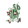
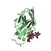
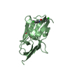
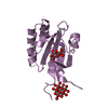
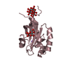
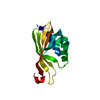
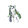
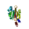
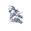
Entry Database : PDB / ID : 5n86Title Crystal structure of FAS1 domain of hyaluronic acid receptor stabilin-2 Stabilin-2 Keywords / / / / Function / homology Function Domain/homology Component
/ / / / / / / / / / / / / / / / / / / / / / / / / / / / / / / / / / / / / / / / / / / / / / / / / / / / / / / / / / / / / Biological species Homo sapiens (human)Method / / / / Resolution : 1.484 Å Authors Twarda-Clapa, A. / Labuzek, B. / Grudnik, P. / Dubin, G. / Holak, T.A. Funding support Organization Grant number Country National Science Centre UMO-2015/17/N/NZ1/00025 National Science Centre UMO-2012/06/A/ST5/00224
Journal : Acta Crystallogr D Struct Biol / Year : 2018Title : Crystal structure of the FAS1 domain of the hyaluronic acid receptor stabilin-2.Authors : Twarda-Clapa, A. / Labuzek, B. / Krzemien, D. / Musielak, B. / Grudnik, P. / Dubin, G. / Holak, T.A. History Deposition Feb 23, 2017 Deposition site / Processing site Revision 1.0 Jun 27, 2018 Provider / Type Revision 1.1 Jul 11, 2018 Group / Database references / Category Item _citation.country / _citation.journal_abbrev ... _citation.country / _citation.journal_abbrev / _citation.journal_id_ASTM / _citation.journal_id_ISSN / _citation.journal_volume / _citation.page_first / _citation.page_last / _citation.pdbx_database_id_PubMed / _citation.title Revision 1.2 Jan 17, 2024 Group / Database references / Refinement descriptionCategory chem_comp_atom / chem_comp_bond ... chem_comp_atom / chem_comp_bond / database_2 / pdbx_initial_refinement_model Item / _database_2.pdbx_database_accession
Show all Show less
 Movie
Movie Controller

 Yorodumi
 Open data
 Basic information
 Components
 Keywords
 Function and homology information
 Homo sapiens (human)
 X-RAY DIFFRACTION /
  Authors
 Poland, 2items
  Citation
 Structure visualization
 Downloads & links
 Other downloads
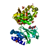


 PDBj
PDBj


 Assembly


 Mass: 18.015 Da / Num. of mol.: 361 / Source method: isolated from a natural source / Formula: H2O
 Sample preparation
 / Beamline: 14.1 / Wavelength: 0.9184 Å
 Processing