 Movie
Movie Controller


+ Open data

- Basic information
| Entry | Database: PDB / ID: 5mqn | ||||||
|---|---|---|---|---|---|---|---|
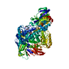
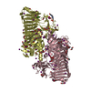
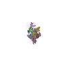
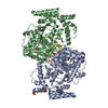
| Title | Glycoside hydrolase BT_0986 | ||||||
 Components Components | Glycosyl hydrolases family 2, sugar binding domain | ||||||
 Keywords Keywords | HYDROLASE / glycoside hydrolase / rhamnosidase / plant pectin / CAZy family 106 | ||||||
| Function / homology | alpha-L-rhamnosidase / Galactose-binding-like domain superfamily / Prokaryotic membrane lipoprotein lipid attachment site profile. / hydrolase activity / DNA binding / metal ion binding / DNA-binding protein / Glycoside hydrolase family 2, sugar binding protein Function and homology information Function and homology information | ||||||
| Biological species |  Bacteroides thetaiotaomicron (bacteria) Bacteroides thetaiotaomicron (bacteria) | ||||||
| Method |  X-RAY DIFFRACTION / SYNCHROTRON / SAD / Resolution: 1.9 Å X-RAY DIFFRACTION / SYNCHROTRON / SAD / Resolution: 1.9 Å | ||||||
 Authors Authors | Basle, A. / Ndeh, D. / Rogowski, A. / Cartmell, A. / Luis, A.S. / Venditto, I. / Labourel, A. / Gilbert, H.J. | ||||||
 Citation Citation | Journal: Nature / Year: 2017 Title: Complex pectin metabolism by gut bacteria reveals novel catalytic functions. Authors: Ndeh, D. / Rogowski, A. / Cartmell, A. / Luis, A.S. / Basle, A. / Gray, J. / Venditto, I. / Briggs, J. / Zhang, X. / Labourel, A. / Terrapon, N. / Buffetto, F. / Nepogodiev, S. / Xiao, Y. / ...Authors: Ndeh, D. / Rogowski, A. / Cartmell, A. / Luis, A.S. / Basle, A. / Gray, J. / Venditto, I. / Briggs, J. / Zhang, X. / Labourel, A. / Terrapon, N. / Buffetto, F. / Nepogodiev, S. / Xiao, Y. / Field, R.A. / Zhu, Y. / O'Neill, M.A. / Urbanowicz, B.R. / York, W.S. / Davies, G.J. / Abbott, D.W. / Ralet, M.C. / Martens, E.C. / Henrissat, B. / Gilbert, H.J. | ||||||
| History |
|
- Structure visualization
| Structure viewer | Molecule: MolmilJmol/JSmol |
|---|
- Downloads & links
-Download
| PDBx/mmCIF format | 5mqn.cif.gz | 441 KB | Display | PDBx/mmCIF format |
|---|---|---|---|---|
| PDB format | pdb5mqn.ent.gz | 356.9 KB | Display | PDB format |
| PDBx/mmJSON format | 5mqn.json.gz | Tree view | PDBx/mmJSON format | |
| Others |  Other downloads Other downloads |
-Validation report
| Summary document | 5mqn_validation.pdf.gz | 423.7 KB | Display | wwPDB validaton report |
|---|---|---|---|---|
| Full document | 5mqn_full_validation.pdf.gz | 428.8 KB | Display | |
| Data in XML | 5mqn_validation.xml.gz | 45.4 KB | Display | |
| Data in CIF | 5mqn_validation.cif.gz | 69.7 KB | Display | |
| Arichive directory | https://data.pdbj.org/pub/pdb/validation_reports/mq/5mqnftp://data.pdbj.org/pub/pdb/validation_reports/mq/5mqn | HTTPS FTP |
-Related structure data
| Related structure data |  5mqmC  5mqoC  5mqrC  5mqsC  5msxC  5msyC  5mt2C  5muiC  5mujC  5mwkC C: citing same article ( |
|---|---|
| Similar structure data |


-Links
 PDBj
PDBj


- Assembly
| Deposited unit | 
| ||||||||
|---|---|---|---|---|---|---|---|---|---|
| 1 |
| ||||||||
| Unit cell |
|
-Components
| #1: Protein | Mass: 125135.859 Da / Num. of mol.: 1 Source method: isolated from a genetically manipulated source Source: (gene. exp.) Bacteroides thetaiotaomicron (bacteria)Gene: Btheta7330_02599 / Plasmid: pet28b / Production host: | ||
|---|---|---|---|
| #2: Chemical |   Mass: 40.078 Da / Num. of mol.: 3 / Source method: obtained synthetically / Formula: Ca Mass: 40.078 Da / Num. of mol.: 3 / Source method: obtained synthetically / Formula: Ca#3: Water | ChemComp-HOH / |  Mass: 18.015 Da / Num. of mol.: 968 / Source method: isolated from a natural source / Formula: H2O Mass: 18.015 Da / Num. of mol.: 968 / Source method: isolated from a natural source / Formula: H2O |
-Experimental details
-Experiment
| Experiment | Method: X-RAY DIFFRACTION / Number of used crystals: 1 |
|---|
- Sample preparation
| Crystal | Density Matthews: 2.63 Å3/Da / Density % sol: 53.26 % |
|---|---|
| Crystal grow | Temperature: 293.15 K / Method: vapor diffusion, sitting drop Details: 15% (w/v) PEG 550 MME, 15% (w/v) PEG 20000, 0.25 M rahmnose, 50mM hepes and 50 mm MOPS pH 7.5 |
-Data collection
| Diffraction | Mean temperature: 100 K |
|---|---|
| Diffraction source | Source: SYNCHROTRON / Site: Diamond  / Beamline: I02 / Wavelength: 0.913 Å / Beamline: I02 / Wavelength: 0.913 Å |
| Detector | Type: DECTRIS PILATUS 6M / Detector: PIXEL / Date: May 24, 2015 |
| Radiation | Protocol: SINGLE WAVELENGTH / Monochromatic (M) / Laue (L): M / Scattering type: x-ray |
| Radiation wavelength | Wavelength: 0.913 Å / Relative weight: 1 |
| Reflection | Resolution: 1.9→49.17 Å / Num. all: 380421 / Num. obs: 101961 / % possible obs: 99.7 % / Observed criterion σ(I): 1.5 / Redundancy: 3.7 % / CC1/2: 0.996 / Rmerge(I) obs: 0.076 / Net I/σ(I): 9.7 |
| Reflection shell | Resolution: 1.9→1.93 Å / Redundancy: 3.2 % / Rmerge(I) obs: 0.653 / Mean I/σ(I) obs: 1.7 / Num. unique all: 4655 / CC1/2: 0.671 / % possible all: 99.1 |
- Processing
| Software |
| ||||||||||||||||||||||||||||||||||||||||||||||||||||||||||||||||||||||||||||||||||||||||||||||||||||||||||||||||||||||||||||||||||||||||||||||||||||||||||||||||||||||||||||||||||||||
|---|---|---|---|---|---|---|---|---|---|---|---|---|---|---|---|---|---|---|---|---|---|---|---|---|---|---|---|---|---|---|---|---|---|---|---|---|---|---|---|---|---|---|---|---|---|---|---|---|---|---|---|---|---|---|---|---|---|---|---|---|---|---|---|---|---|---|---|---|---|---|---|---|---|---|---|---|---|---|---|---|---|---|---|---|---|---|---|---|---|---|---|---|---|---|---|---|---|---|---|---|---|---|---|---|---|---|---|---|---|---|---|---|---|---|---|---|---|---|---|---|---|---|---|---|---|---|---|---|---|---|---|---|---|---|---|---|---|---|---|---|---|---|---|---|---|---|---|---|---|---|---|---|---|---|---|---|---|---|---|---|---|---|---|---|---|---|---|---|---|---|---|---|---|---|---|---|---|---|---|---|---|---|---|
| Refinement | Method to determine structure: SAD / Resolution: 1.9→49.17 Å / Cor.coef. Fo:Fc: 0.96 / Cor.coef. Fo:Fc free: 0.942 / SU B: 6.083 / SU ML: 0.095 / Cross valid method: THROUGHOUT / ESU R: 0.132 / ESU R Free: 0.123 / Details: HYDROGENS HAVE BEEN ADDED IN THE RIDING POSITIONS
| ||||||||||||||||||||||||||||||||||||||||||||||||||||||||||||||||||||||||||||||||||||||||||||||||||||||||||||||||||||||||||||||||||||||||||||||||||||||||||||||||||||||||||||||||||||||
| Solvent computation | Ion probe radii: 0.8 Å / Shrinkage radii: 0.8 Å / VDW probe radii: 1.2 Å | ||||||||||||||||||||||||||||||||||||||||||||||||||||||||||||||||||||||||||||||||||||||||||||||||||||||||||||||||||||||||||||||||||||||||||||||||||||||||||||||||||||||||||||||||||||||
| Displacement parameters | Biso mean: 30.217 Å2
| ||||||||||||||||||||||||||||||||||||||||||||||||||||||||||||||||||||||||||||||||||||||||||||||||||||||||||||||||||||||||||||||||||||||||||||||||||||||||||||||||||||||||||||||||||||||
| Refinement step | Cycle: 1 / Resolution: 1.9→49.17 Å
| ||||||||||||||||||||||||||||||||||||||||||||||||||||||||||||||||||||||||||||||||||||||||||||||||||||||||||||||||||||||||||||||||||||||||||||||||||||||||||||||||||||||||||||||||||||||
| Refine LS restraints |
|