 Movie
Movie Controller

[English] 日本語
 Yorodumi
- PDB-7jwf: Crystal structure of PdGH110B D344N in complex with alpha-(1,3)-g... -
+ Open data

- Basic information
| Entry | Database: PDB / ID: 7jwf | ||||||
|---|---|---|---|---|---|---|---|
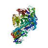
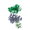
| Title | Crystal structure of PdGH110B D344N in complex with alpha-(1,3)-galactobiose | ||||||
 Components Components | Glycoside hydrolase family 110 | ||||||
 Keywords Keywords | HYDROLASE / beta helix | ||||||
| Function / homology | ACETATE ION / IODIDE ION / MALONIC ACID / MALONATE ION / D-MALATE Function and homology information Function and homology information | ||||||
| Biological species |  Pseudoalteromonas distincta (bacteria) Pseudoalteromonas distincta (bacteria) | ||||||
| Method |  X-RAY DIFFRACTION / MOLECULAR REPLACEMENT / Resolution: 2.187 Å X-RAY DIFFRACTION / MOLECULAR REPLACEMENT / Resolution: 2.187 Å | ||||||
 Authors Authors | Hettle, A.G. / Boraston, A.B. | ||||||
| Funding support | 1items
| ||||||
 Citation Citation | Journal: J.Biol.Chem. / Year: 2020 Title: The structure of a family 110 glycoside hydrolase provides insight into the hydrolysis of alpha-1,3-galactosidic linkages in lambda-carrageenan and blood group antigens. Authors: McGuire, B.E. / Hettle, A.G. / Vickers, C. / King, D.T. / Vocadlo, D.J. / Boraston, A.B. | ||||||
| History |
|
- Structure visualization
| Structure viewer | Molecule: MolmilJmol/JSmol |
|---|
- Downloads & links
-Download
| PDBx/mmCIF format | 7jwf.cif.gz | 516.4 KB | Display | PDBx/mmCIF format |
|---|---|---|---|---|
| PDB format | pdb7jwf.ent.gz | 412.6 KB | Display | PDB format |
| PDBx/mmJSON format | 7jwf.json.gz | Tree view | PDBx/mmJSON format | |
| Others |  Other downloads Other downloads |
-Validation report
| Arichive directory | https://data.pdbj.org/pub/pdb/validation_reports/jw/7jwfftp://data.pdbj.org/pub/pdb/validation_reports/jw/7jwf | HTTPS FTP |
|---|
-Related structure data
| Related structure data |  7jw4SC S: Starting model for refinement C: citing same article ( |
|---|---|
| Similar structure data |

-Links
 PDBj
PDBj


- Assembly
| Deposited unit | 
| ||||||||
|---|---|---|---|---|---|---|---|---|---|
| 1 | 
| ||||||||
| 2 | 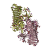
| ||||||||
| Unit cell |
|
-Components
-Protein / Sugars , 2 types, 8 molecules ABCD
| #1: Protein | Mass: 69845.688 Da / Num. of mol.: 4 Source method: isolated from a genetically manipulated source Source: (gene. exp.) Pseudoalteromonas distincta (bacteria) / Plasmid: pET28a / Production host: #2: Polysaccharide | alpha-D-galactopyranose-(1-3)-beta-D-galactopyranose Source method: isolated from a genetically manipulated source |
|---|
-Non-polymers , 11 types, 1853 molecules 


| #3: Chemical | ChemComp-MLT /  Mass: 134.087 Da / Num. of mol.: 4 / Source method: obtained synthetically / Formula: C4H6O5 Mass: 134.087 Da / Num. of mol.: 4 / Source method: obtained synthetically / Formula: C4H6O5#4: Chemical | ChemComp-EDO /  Mass: 62.068 Da / Num. of mol.: 50 / Source method: obtained synthetically / Formula: C2H6O2 Mass: 62.068 Da / Num. of mol.: 50 / Source method: obtained synthetically / Formula: C2H6O2#5: Chemical | ChemComp-IOD /  Mass: 126.904 Da / Num. of mol.: 119 / Source method: obtained synthetically / Formula: I Mass: 126.904 Da / Num. of mol.: 119 / Source method: obtained synthetically / Formula: I#6: Chemical |  Mass: 194.226 Da / Num. of mol.: 2 / Source method: obtained synthetically / Formula: C8H18O5 / Comment: precipitant*YM Mass: 194.226 Da / Num. of mol.: 2 / Source method: obtained synthetically / Formula: C8H18O5 / Comment: precipitant*YM#7: Chemical | ChemComp-CA / |  Mass: 40.078 Da / Num. of mol.: 1 / Source method: obtained synthetically / Formula: Ca Mass: 40.078 Da / Num. of mol.: 1 / Source method: obtained synthetically / Formula: Ca#8: Chemical |  Mass: 59.044 Da / Num. of mol.: 3 / Source method: obtained synthetically / Formula: C2H3O2 Mass: 59.044 Da / Num. of mol.: 3 / Source method: obtained synthetically / Formula: C2H3O2#9: Chemical | ChemComp-CL /  Mass: 35.453 Da / Num. of mol.: 10 / Source method: obtained synthetically / Formula: Cl Mass: 35.453 Da / Num. of mol.: 10 / Source method: obtained synthetically / Formula: Cl#10: Chemical | ChemComp-MLA / |  Mass: 104.061 Da / Num. of mol.: 1 / Source method: obtained synthetically / Formula: C3H4O4 Mass: 104.061 Da / Num. of mol.: 1 / Source method: obtained synthetically / Formula: C3H4O4#11: Chemical | ChemComp-MLI / |  Mass: 102.046 Da / Num. of mol.: 1 / Source method: obtained synthetically / Formula: C3H2O4 Mass: 102.046 Da / Num. of mol.: 1 / Source method: obtained synthetically / Formula: C3H2O4#12: Chemical | ChemComp-EPE / |  Mass: 238.305 Da / Num. of mol.: 1 / Source method: obtained synthetically / Formula: C8H18N2O4S / Comment: pH buffer*YM Mass: 238.305 Da / Num. of mol.: 1 / Source method: obtained synthetically / Formula: C8H18N2O4S / Comment: pH buffer*YM#13: Water | ChemComp-HOH / | Mass: 18.015 Da / Num. of mol.: 1661 / Source method: isolated from a natural source / Formula: H2O |
|---|
-Details
| Has ligand of interest | Y |
|---|
-Experimental details
-Experiment
| Experiment | Method: X-RAY DIFFRACTION / Number of used crystals: 1 |
|---|
- Sample preparation
| Crystal | Density Matthews: 3.24 Å3/Da / Density % sol: 62.04 % |
|---|---|
| Crystal grow | Temperature: 291 K / Method: vapor diffusion, sitting drop / pH: 7.5 / Details: PEG 3350, HEPES, NaI, Tacsimate |
-Data collection
| Diffraction | Mean temperature: 100 K / Serial crystal experiment: N |
|---|---|
| Diffraction source | Source: ROTATING ANODE / Type: RIGAKU MICROMAX-002 / Wavelength: 1.514 Å |
| Detector | Type: DECTRIS PILATUS 200K / Detector: PIXEL / Date: Dec 2, 2016 |
| Radiation | Protocol: SINGLE WAVELENGTH / Monochromatic (M) / Laue (L): M / Scattering type: x-ray |
| Radiation wavelength | Wavelength: 1.514 Å / Relative weight: 1 |
| Reflection | Resolution: 2.187→30 Å / Num. obs: 174470 / % possible obs: 98.6 % / Redundancy: 3.6 % / CC1/2: 0.991 / Net I/σ(I): 7.1 |
| Reflection shell | Resolution: 2.2→2.24 Å / Num. unique obs: 7427 / CC1/2: 0.833 |
- Processing
| Software |
| ||||||||||||||||||||||||||||||||||||||||||||||||||||||||||||||||||||||||||||||||||||||||||||||||||||||||||||||||||||||||||||||||||||||||||||||||||||||||||||||||||||||||||||||||||||||||||
|---|---|---|---|---|---|---|---|---|---|---|---|---|---|---|---|---|---|---|---|---|---|---|---|---|---|---|---|---|---|---|---|---|---|---|---|---|---|---|---|---|---|---|---|---|---|---|---|---|---|---|---|---|---|---|---|---|---|---|---|---|---|---|---|---|---|---|---|---|---|---|---|---|---|---|---|---|---|---|---|---|---|---|---|---|---|---|---|---|---|---|---|---|---|---|---|---|---|---|---|---|---|---|---|---|---|---|---|---|---|---|---|---|---|---|---|---|---|---|---|---|---|---|---|---|---|---|---|---|---|---|---|---|---|---|---|---|---|---|---|---|---|---|---|---|---|---|---|---|---|---|---|---|---|---|---|---|---|---|---|---|---|---|---|---|---|---|---|---|---|---|---|---|---|---|---|---|---|---|---|---|---|---|---|---|---|---|---|
| Refinement | Method to determine structure: MOLECULAR REPLACEMENT Starting model: 7JW4 Resolution: 2.187→29.759 Å / SU ML: 0.27 / Cross valid method: THROUGHOUT / σ(F): 1.33 / Phase error: 26.07 / Stereochemistry target values: ML
| ||||||||||||||||||||||||||||||||||||||||||||||||||||||||||||||||||||||||||||||||||||||||||||||||||||||||||||||||||||||||||||||||||||||||||||||||||||||||||||||||||||||||||||||||||||||||||
| Solvent computation | Shrinkage radii: 0.9 Å / VDW probe radii: 1.11 Å / Solvent model: FLAT BULK SOLVENT MODEL | ||||||||||||||||||||||||||||||||||||||||||||||||||||||||||||||||||||||||||||||||||||||||||||||||||||||||||||||||||||||||||||||||||||||||||||||||||||||||||||||||||||||||||||||||||||||||||
| Displacement parameters | Biso max: 135.47 Å2 / Biso mean: 34.902 Å2 / Biso min: 18.51 Å2 | ||||||||||||||||||||||||||||||||||||||||||||||||||||||||||||||||||||||||||||||||||||||||||||||||||||||||||||||||||||||||||||||||||||||||||||||||||||||||||||||||||||||||||||||||||||||||||
| Refinement step | Cycle: final / Resolution: 2.187→29.759 Å
| ||||||||||||||||||||||||||||||||||||||||||||||||||||||||||||||||||||||||||||||||||||||||||||||||||||||||||||||||||||||||||||||||||||||||||||||||||||||||||||||||||||||||||||||||||||||||||
| LS refinement shell | Refine-ID: X-RAY DIFFRACTION / Rfactor Rfree error: 0
|
