 Movie
Movie Controller


+ Open data

- Basic information
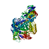
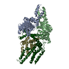
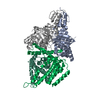
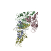
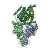
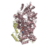
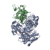
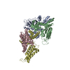
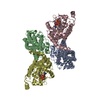
| Entry | Database: PDB / ID: 4rfs | ||||||
|---|---|---|---|---|---|---|---|
| Title | Structure of a pantothenate energy coupling factor transporter | ||||||
 Components Components |
| ||||||
 Keywords Keywords | HYDROLASE / TRANSPORT PROTEIN / Transporter / ECF | ||||||
| Function / homology |  Function and homology information Function and homology informationTranslocases / ATPase-coupled transmembrane transporter activity / transmembrane transporter activity / ATP-binding cassette (ABC) transporter complex / ATP hydrolysis activity / ATP binding / membrane / plasma membrane Similarity search - Function | ||||||
| Biological species |  Lactobacillus brevis (bacteria) Lactobacillus brevis (bacteria) | ||||||
| Method |  X-RAY DIFFRACTION / SYNCHROTRON / MOLECULAR REPLACEMENT / Resolution: 3.232 Å X-RAY DIFFRACTION / SYNCHROTRON / MOLECULAR REPLACEMENT / Resolution: 3.232 Å | ||||||
 Authors Authors | Zhang, M. / Bao, Z. / Zhao, Q. / Guo, H. / Xu, K. / Zhang, P. | ||||||
 Citation Citation | Journal: Proc.Natl.Acad.Sci.USA / Year: 2014 Title: Structure of a pantothenate transporter and implications for ECF module sharing and energy coupling of group II ECF transporters. Authors: Zhang, M. / Bao, Z. / Zhao, Q. / Guo, H. / Xu, K. / Wang, C. / Zhang, P. | ||||||
| History |
|
- Structure visualization
| Structure viewer | Molecule: MolmilJmol/JSmol |
|---|
- Downloads & links
-Download
| PDBx/mmCIF format | 4rfs.cif.gz | 394.3 KB | Display | PDBx/mmCIF format |
|---|---|---|---|---|
| PDB format | pdb4rfs.ent.gz | 324.6 KB | Display | PDB format |
| PDBx/mmJSON format | 4rfs.json.gz | Tree view | PDBx/mmJSON format | |
| Others |  Other downloads Other downloads |
-Validation report
| Arichive directory | https://data.pdbj.org/pub/pdb/validation_reports/rf/4rfsftp://data.pdbj.org/pub/pdb/validation_reports/rf/4rfs | HTTPS FTP |
|---|
-Related structure data
| Similar structure data |
|---|

-Links
 PDBj
PDBj

- Assembly
| Deposited unit | 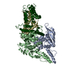
| ||||||||
|---|---|---|---|---|---|---|---|---|---|
| 1 |
| ||||||||
| Unit cell |
|
-Components
| #1: Protein | Mass: 32176.533 Da / Num. of mol.: 1 Source method: isolated from a genetically manipulated source Source: (gene. exp.) Lactobacillus brevis (bacteria) / Gene: ecfA2 / Production host: References: UniProt: Q03PY6, Hydrolases; Acting on acid anhydrides; Acting on acid anhydrides to catalyse transmembrane movement of substances |
|---|---|
| #2: Protein | Mass: 30550.432 Da / Num. of mol.: 1 Source method: isolated from a genetically manipulated source Source: (gene. exp.) Lactobacillus brevis (bacteria) / Gene: ecfA1 / Production host: References: UniProt: Q03PY5, Hydrolases; Acting on acid anhydrides; Acting on acid anhydrides to catalyse transmembrane movement of substances |
| #3: Protein | Mass: 22209.598 Da / Num. of mol.: 1 Source method: isolated from a genetically manipulated source Source: (gene. exp.) Lactobacillus brevis (bacteria) / Gene: LVIS_0658 / Production host: |
| #4: Protein | Mass: 31939.779 Da / Num. of mol.: 1 Source method: isolated from a genetically manipulated source Source: (gene. exp.) Lactobacillus brevis (bacteria) / Gene: ecfT / Production host: |
-Experimental details
-Experiment
| Experiment | Method: X-RAY DIFFRACTION / Number of used crystals: 1 |
|---|
- Sample preparation
| Crystal | Density Matthews: 3.81 Å3/Da / Density % sol: 67.68 % |
|---|---|
| Crystal grow | Temperature: 293 K / Method: vapor diffusion / pH: 8.4 Details: 0.1 M Tris, 20% PEG2000, 15% glycerol, 0.2M MgCl2, pH 8.4, VAPOR DIFFUSION, temperature 293K |
-Data collection
| Diffraction | Mean temperature: 100 K |
|---|---|
| Diffraction source | Source: SYNCHROTRON / Site: SSRF  / Beamline: BL17U / Wavelength: 0.97913 Å / Beamline: BL17U / Wavelength: 0.97913 Å |
| Detector | Type: ADSC QUANTUM 315r / Detector: CCD / Date: Jan 20, 2013 |
| Radiation | Protocol: SINGLE WAVELENGTH / Monochromatic (M) / Laue (L): M / Scattering type: x-ray |
| Radiation wavelength | Wavelength: 0.97913 Å / Relative weight: 1 |
| Reflection | Resolution: 3.25→37.5 Å / Num. obs: 28312 / % possible obs: 97.6 % / Redundancy: 4.9 % / Rmerge(I) obs: 0.08 / Net I/σ(I): 21.4 |
| Reflection shell | Resolution: 3.25→3.37 Å / % possible obs: 97.6 % / Redundancy: 4.8 % / Rmerge(I) obs: 0.884 / Mean I/σ(I) obs: 1.9 / % possible all: 97.6 |
- Processing
| Software |
| |||||||||||||||||||||||||||||||||||||||||||||||||||||||||||||||||||||||||||||
|---|---|---|---|---|---|---|---|---|---|---|---|---|---|---|---|---|---|---|---|---|---|---|---|---|---|---|---|---|---|---|---|---|---|---|---|---|---|---|---|---|---|---|---|---|---|---|---|---|---|---|---|---|---|---|---|---|---|---|---|---|---|---|---|---|---|---|---|---|---|---|---|---|---|---|---|---|---|---|
| Refinement | Method to determine structure: MOLECULAR REPLACEMENT / Resolution: 3.232→37.5 Å / SU ML: 1.24 / σ(F): 1.34 / Phase error: 30.08 / Stereochemistry target values: ML
| |||||||||||||||||||||||||||||||||||||||||||||||||||||||||||||||||||||||||||||
| Solvent computation | Shrinkage radii: 0.9 Å / VDW probe radii: 1.11 Å / Solvent model: FLAT BULK SOLVENT MODEL / Bsol: 40.77 Å2 / ksol: 0.29 e/Å3 | |||||||||||||||||||||||||||||||||||||||||||||||||||||||||||||||||||||||||||||
| Displacement parameters |
| |||||||||||||||||||||||||||||||||||||||||||||||||||||||||||||||||||||||||||||
| Refinement step | Cycle: LAST / Resolution: 3.232→37.5 Å
| |||||||||||||||||||||||||||||||||||||||||||||||||||||||||||||||||||||||||||||
| Refine LS restraints |
| |||||||||||||||||||||||||||||||||||||||||||||||||||||||||||||||||||||||||||||
| LS refinement shell |
| |||||||||||||||||||||||||||||||||||||||||||||||||||||||||||||||||||||||||||||
| Refinement TLS params. | Method: refined / Origin x: 15.838 Å / Origin y: 170.9193 Å / Origin z: 186.2089 Å
| |||||||||||||||||||||||||||||||||||||||||||||||||||||||||||||||||||||||||||||
| Refinement TLS group | Selection details: all |
