[English] 日本語
Yorodumi
- PDB-5x3x: 2.8A resolution structure of a cobalt energy-coupling factor tran... -

+
Open data


ID or keywords:

Loading...

-
Basic information

Entry
Database: PDB / ID: 5x3x
Title
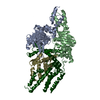
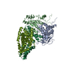
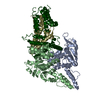
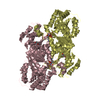
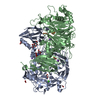
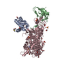
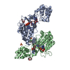
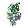
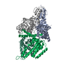
2.8A resolution structure of a cobalt energy-coupling factor transporter-CbiMQO
Components
  • Cobalt ABC transporter ATP-binding protein
  • Cobalt transport protein CbiM
  • Uncharacterized protein CbiQ
KeywordsTRANSPORT PROTEIN / Complex / ECF transporter
Function / homology
Function and homology information


Translocases; Catalysing the translocation of inorganic cations; Linked to the hydrolysis of a nucleoside triphosphate / cobalt ion transport / cobalt ion transmembrane transporter activity / cobalamin biosynthetic process / plasma membrane => GO:0005886 / ATP-binding cassette (ABC) transporter complex / ATP hydrolysis activity / ATP binding
Similarity search - Function
Cobalt ECF transporter T component CbiQ / Cobalamin (vitamin B12) transport protein CbiM / Cobalt transport protein ATP-binding subunit / Metal transport protein CbiM/NikMN / Cobalt uptake substrate-specific transmembrane region / ABC/ECF transporter, transmembrane component / Cobalt transport protein / ABC transporter, CbiO/EcfA subunit / ABC transporter-like, conserved site / ABC transporters family signature. ...Cobalt ECF transporter T component CbiQ / Cobalamin (vitamin B12) transport protein CbiM / Cobalt transport protein ATP-binding subunit / Metal transport protein CbiM/NikMN / Cobalt uptake substrate-specific transmembrane region / ABC/ECF transporter, transmembrane component / Cobalt transport protein / ABC transporter, CbiO/EcfA subunit / ABC transporter-like, conserved site / ABC transporters family signature. / ABC transporter / ABC transporter-like, ATP-binding domain / ATP-binding cassette, ABC transporter-type domain profile. / P-loop containing nucleotide triphosphate hydrolases / ATPases associated with a variety of cellular activities / AAA+ ATPase domain / P-loop containing nucleoside triphosphate hydrolase / Rossmann fold / 3-Layer(aba) Sandwich / Alpha Beta
Similarity search - Domain/homology
Cobalt transport protein CbiM / Cobalt transport protein CbiQ / Cobalt transport protein CbiM / Cobalt import ATP-binding protein CbiO
Similarity search - Component
Biological speciesRhodobacter capsulatus (bacteria)
MethodX-RAY DIFFRACTION / SYNCHROTRON / SAD / Resolution: 2.788 Å
AuthorsBao, Z. / Qi, X. / Wang, J. / Zhang, P.
CitationJournal: Cell Res. / Year: 2017
Title: Structure and mechanism of a group-I cobalt energy coupling factor transporter
Authors: Bao, Z. / Qi, X. / Hong, S. / Xu, K. / He, F. / Zhang, M. / Chen, J. / Chao, D. / Zhao, W. / Li, D. / Wang, J. / Zhang, P.
History
DepositionFeb 9, 2017Deposition site: PDBJ / Processing site: PDBJ
Revision 1.0Apr 5, 2017Provider: repository / Type: Initial release
Revision 1.1Apr 19, 2017Group: Structure summary
Revision 1.2May 17, 2017Group: Database references
Revision 1.3Oct 4, 2017Group: Data collection / Category: diffrn_detector / Item: _diffrn_detector.detector
Revision 1.4Mar 27, 2024Group: Data collection / Database references / Category: chem_comp_atom / chem_comp_bond / database_2
Item: _database_2.pdbx_DOI / _database_2.pdbx_database_accession

-
Structure visualization

Structure viewerMolecule:
MolmilJmol/JSmol

Downloads & links

-
Assembly

Deposited unit
A: Cobalt ABC transporter ATP-binding protein
B: Cobalt ABC transporter ATP-binding protein
M: Cobalt transport protein CbiM
Q: Uncharacterized protein CbiQ
a: Cobalt ABC transporter ATP-binding protein
b: Cobalt ABC transporter ATP-binding protein
m: Cobalt transport protein CbiM
q: Uncharacterized protein CbiQ


Theoretical massNumber of molelcules
Total (without water)214,7518
Polymers214,7518
Non-polymers00
Water2,630146
1
A: Cobalt ABC transporter ATP-binding protein
B: Cobalt ABC transporter ATP-binding protein
M: Cobalt transport protein CbiM
Q: Uncharacterized protein CbiQ


Theoretical massNumber of molelcules
Total (without water)107,3764
Polymers107,3764
Non-polymers00
Water724
TypeNameSymmetry operationNumber
identity operation1_555x,y,z1
2
a: Cobalt ABC transporter ATP-binding protein
b: Cobalt ABC transporter ATP-binding protein
m: Cobalt transport protein CbiM
q: Uncharacterized protein CbiQ


Theoretical massNumber of molelcules
Total (without water)107,3764
Polymers107,3764
Non-polymers00
Water724
TypeNameSymmetry operationNumber
identity operation1_555x,y,z1
Unit cell
Length a, b, c (Å)63.676, 220.564, 301.277
Angle α, β, γ (deg.)90.00, 90.00, 90.00
Int Tables number20
Space group name H-MC2221

-
Components

#1: Protein
Cobalt ABC transporter ATP-binding protein / CbiO


Mass: 29464.053 Da / Num. of mol.: 4
Source method: isolated from a genetically manipulated source
Source: (gene. exp.) Rhodobacter capsulatus (bacteria) / Production host: Escherichia coli (E. coli) / References: UniProt: O68106*PLUS
#2: Protein Cobalt transport protein CbiM / Energy-coupling factor transporter probable substrate-capture protein CbiM / ECF transporter S component CbiM


Mass: 22554.672 Da / Num. of mol.: 2
Source method: isolated from a genetically manipulated source
Source: (gene. exp.) Rhodobacter capsulatus (bacteria) / Gene: cbiM / Production host: Escherichia coli (E. coli) / References: UniProt: A0A0Q0R232, UniProt: D5AUZ9*PLUS
#3: Protein Uncharacterized protein CbiQ


Mass: 25892.801 Da / Num. of mol.: 2
Source method: isolated from a genetically manipulated source
Source: (gene. exp.) Rhodobacter capsulatus (bacteria) / Production host: Escherichia coli (E. coli) / References: UniProt: D5AUZ7*PLUS
#4: Water ChemComp-HOH / water / Water


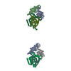
Mass: 18.015 Da / Num. of mol.: 146 / Source method: isolated from a natural source / Formula: H2O

-
Experimental details

-
Experiment

ExperimentMethod: X-RAY DIFFRACTION / Number of used crystals: 1

-
Sample preparation

CrystalDensity Matthews: 2.46 Å3/Da / Density % sol: 50.06 %
Crystal growTemperature: 293 K / Method: vapor diffusion, hanging drop / Details: 1M ammonium sulfate, 0.1M ADA, pH6.5

-
Data collection

DiffractionMean temperature: 100 K
Diffraction sourceSource: SYNCHROTRON / Site: SSRF / Beamline: BL19U1 / Wavelength: 0.978 Å
DetectorType: MAR555 FLAT PANEL / Detector: IMAGE PLATE / Date: Nov 10, 2015
RadiationProtocol: SINGLE WAVELENGTH / Monochromatic (M) / Laue (L): M / Scattering type: x-ray
Radiation wavelengthWavelength: 0.978 Å / Relative weight: 1
ReflectionResolution: 2.78→50 Å / Num. obs: 51793 / % possible obs: 96.4 % / Redundancy: 3.6 % / Rmerge(I) obs: 0.113 / Net I/σ(I): 7.6

-
Processing

Software
NameVersionClassification
PHENIX1.9_1692refinement
HKL-3000data collection
HKL-3000data processing
PHENIXphasing
RefinementMethod to determine structure: SAD / Resolution: 2.788→48.334 Å / SU ML: 0.34 / Cross valid method: FREE R-VALUE / σ(F): 1.34 / Phase error: 28.49 / Stereochemistry target values: ML
RfactorNum. reflection% reflection
Rfree0.2486 2376 5.1 %
Rwork0.2141 --
obs0.2159 46628 87 %
Solvent computationShrinkage radii: 0.9 Å / VDW probe radii: 1.11 Å / Solvent model: FLAT BULK SOLVENT MODEL
Refinement stepCycle: LAST / Resolution: 2.788→48.334 Å
ProteinNucleic acidLigandSolventTotal
Num. atoms14820 0 0 146 14966
Refine LS restraints
Refine-IDTypeDev idealNumber
X-RAY DIFFRACTIONf_bond_d0.01515036
X-RAY DIFFRACTIONf_angle_d1.77120480
X-RAY DIFFRACTIONf_dihedral_angle_d15.495368
X-RAY DIFFRACTIONf_chiral_restr0.0912522
X-RAY DIFFRACTIONf_plane_restr0.0132610
LS refinement shell
Resolution (Å)Rfactor RfreeNum. reflection RfreeRfactor RworkNum. reflection RworkRefine-ID% reflection obs (%)
2.7882-2.84510.278460.2496833X-RAY DIFFRACTION28
2.8451-2.9070.3146780.26841467X-RAY DIFFRACTION50
2.907-2.97460.3113980.25951828X-RAY DIFFRACTION62
2.9746-3.04890.33431100.25862223X-RAY DIFFRACTION75
3.0489-3.13140.27941420.26142540X-RAY DIFFRACTION86
3.1314-3.22350.2881530.24582796X-RAY DIFFRACTION94
3.2235-3.32750.27051660.23222855X-RAY DIFFRACTION97
3.3275-3.44640.27771610.22682943X-RAY DIFFRACTION99
3.4464-3.58440.25511790.21242950X-RAY DIFFRACTION99
3.5844-3.74740.25021410.21122970X-RAY DIFFRACTION99
3.7474-3.94490.25611370.20482999X-RAY DIFFRACTION100
3.9449-4.19190.22321390.19012948X-RAY DIFFRACTION99
4.1919-4.51540.23061750.18032967X-RAY DIFFRACTION99
4.5154-4.96940.20461460.18762969X-RAY DIFFRACTION98
4.9694-5.68750.23911580.22042978X-RAY DIFFRACTION98
5.6875-7.16190.25831660.24092999X-RAY DIFFRACTION98
7.1619-48.34160.22691810.2052987X-RAY DIFFRACTION94
Refinement TLS params.Method: refined / Origin x: 87.436 Å / Origin y: 134.2336 Å / Origin z: 413.0794 Å
111213212223313233
T0.2639 Å20.0121 Å2-0.0344 Å2-0.2741 Å2-0.0227 Å2--0.2624 Å2
L-0.0266 °2-0.0058 °2-0.0103 °2-0.0487 °20.1703 °2--0.4473 °2
S0.014 Å °-0.0052 Å °0.0118 Å °0.0372 Å °0.0205 Å °-0.0304 Å °0.0618 Å °0.0331 Å °-0.0529 Å °
Refinement TLS groupSelection details: all

+
About Yorodumi

-
News

-
Feb 9, 2022. New format data for meta-information of EMDB entries

New format data for meta-information of EMDB entries

  • Version 3 of the EMDB header file is now the official format.
  • The previous official version 1.9 will be removed from the archive.

Related info.:EMDB header

External links:wwPDB to switch to version 3 of the EMDB data model

-
Aug 12, 2020. Covid-19 info

Covid-19 info

URL: https://pdbj.org/emnavi/covid19.php

New page: Covid-19 featured information page in EM Navigator.

Related info.:Covid-19 info / Mar 5, 2020. Novel coronavirus structure data

+
Mar 5, 2020. Novel coronavirus structure data

Novel coronavirus structure data

Related info.:Yorodumi Speices / Aug 12, 2020. Covid-19 info

External links:COVID-19 featured content - PDBj / Molecule of the Month (242):Coronavirus Proteases

+
Jan 31, 2019. EMDB accession codes are about to change! (news from PDBe EMDB page)

EMDB accession codes are about to change! (news from PDBe EMDB page)

  • The allocation of 4 digits for EMDB accession codes will soon come to an end. Whilst these codes will remain in use, new EMDB accession codes will include an additional digit and will expand incrementally as the available range of codes is exhausted. The current 4-digit format prefixed with “EMD-” (i.e. EMD-XXXX) will advance to a 5-digit format (i.e. EMD-XXXXX), and so on. It is currently estimated that the 4-digit codes will be depleted around Spring 2019, at which point the 5-digit format will come into force.
  • The EM Navigator/Yorodumi systems omit the EMD- prefix.

Related info.:Q: What is EMD? / ID/Accession-code notation in Yorodumi/EM Navigator

External links:EMDB Accession Codes are Changing Soon! / Contact to PDBj

+
Jul 12, 2017. Major update of PDB

Major update of PDB

  • wwPDB released updated PDB data conforming to the new PDBx/mmCIF dictionary.
  • This is a major update changing the version number from 4 to 5, and with Remediation, in which all the entries are updated.
  • In this update, many items about electron microscopy experimental information are reorganized (e.g. em_software).
  • Now, EM Navigator and Yorodumi are based on the updated data.

External links:wwPDB Remediation / Enriched Model Files Conforming to OneDep Data Standards Now Available in the PDB FTP Archive

-
Yorodumi

Thousand views of thousand structures

  • Yorodumi is a browser for structure data from EMDB, PDB, SASBDB, etc.
  • This page is also the successor to EM Navigator detail page, and also detail information page/front-end page for Omokage search.
  • The word "yorodu" (or yorozu) is an old Japanese word meaning "ten thousand". "mi" (miru) is to see.

Related info.:EMDB / PDB / SASBDB / Comparison of 3 databanks / Yorodumi Search / Aug 31, 2016. New EM Navigator & Yorodumi / Yorodumi Papers / Jmol/JSmol / Function and homology information / Changes in new EM Navigator and Yorodumi

Read more