[English] 日本語
Yorodumi
- PDB-5mh6: D-2-hydroxyacid dehydrogenases (D2-HDH) from Haloferax mediterran... -

+
Open data


ID or keywords:

Loading...

-
Basic information

Entry
Database: PDB / ID: 5mh6
TitleD-2-hydroxyacid dehydrogenases (D2-HDH) from Haloferax mediterranei in complex with 2-ketohexanoic acid and NAD+ (1.35 A resolution)
ComponentsD-2-hydroxyacid dehydrogenase
KeywordsOXIDOREDUCTASE / D-2-hydroxyacid dehydrogenase / halophile / chiral specificity / reaction mechanism
Function / homology
Function and homology information


NADH binding / carboxylic acid binding / carboxylic acid metabolic process / Oxidoreductases; Acting on the CH-OH group of donors; With NAD+ or NADP+ as acceptor / oxidoreductase activity, acting on the CH-OH group of donors, NAD or NADP as acceptor / NADPH binding
Similarity search - Function
D-isomer specific 2-hydroxyacid dehydrogenase, NAD-binding domain / D-isomer specific 2-hydroxyacid dehydrogenase, NAD binding domain / NAD(P)-binding Rossmann-like Domain / NAD(P)-binding domain superfamily / Rossmann fold / 3-Layer(aba) Sandwich / Alpha Beta
Similarity search - Domain/homology
2-Ketohexanoic acid / NICOTINAMIDE-ADENINE-DINUCLEOTIDE / D-2-hydroxyacid dehydrogenase
Similarity search - Component
Biological speciesHaloferax mediterranei ATCC 33500 (archaea)
MethodX-RAY DIFFRACTION / SYNCHROTRON / MOLECULAR REPLACEMENT / Resolution: 1.35 Å
AuthorsBisson, C. / Baker, P.J. / Domenech Perez, J. / Pramanpol, N. / Harding, S.E. / Rice, D.W. / Ferrer, J.
Funding support United Kingdom, Spain, 4items
OrganizationGrant numberCountry
Biotechnology and Biological Sciences Research Council United Kingdom
Wellcome Trust United Kingdom
Wolfson Foundation United Kingdom
University of AlicanteVIGROB046 Spain
CitationJournal: To Be Published
Title: Productive ternary complexes of D-2-hydroxyacid dehydrogenase provide insights into the chiral specificity of its reaction mechanism
Authors: Domenech Perez, J. / Pramanpol, N. / Baker, P.J. / Bisson, C. / Harding, S.E. / Rice, D.W. / Ferrer, J.
History
DepositionNov 23, 2016Deposition site: PDBE / Processing site: PDBE
Revision 1.0Jun 6, 2018Provider: repository / Type: Initial release
Revision 2.0Jun 19, 2024Group: Atomic model / Data collection ...Atomic model / Data collection / Database references / Derived calculations / Source and taxonomy
Category: atom_site / atom_site_anisotrop ...atom_site / atom_site_anisotrop / chem_comp_atom / chem_comp_bond / database_2 / entity_src_gen / pdbx_struct_conn_angle / struct_conn
Item: _atom_site.auth_atom_id / _atom_site.label_atom_id ..._atom_site.auth_atom_id / _atom_site.label_atom_id / _atom_site_anisotrop.pdbx_auth_atom_id / _atom_site_anisotrop.pdbx_label_atom_id / _database_2.pdbx_DOI / _database_2.pdbx_database_accession / _entity_src_gen.pdbx_gene_src_scientific_name / _pdbx_struct_conn_angle.ptnr1_auth_asym_id / _pdbx_struct_conn_angle.ptnr1_auth_comp_id / _pdbx_struct_conn_angle.ptnr1_auth_seq_id / _pdbx_struct_conn_angle.ptnr1_label_alt_id / _pdbx_struct_conn_angle.ptnr1_label_asym_id / _pdbx_struct_conn_angle.ptnr1_label_atom_id / _pdbx_struct_conn_angle.ptnr1_label_comp_id / _pdbx_struct_conn_angle.ptnr1_label_seq_id / _pdbx_struct_conn_angle.ptnr1_symmetry / _pdbx_struct_conn_angle.ptnr2_auth_asym_id / _pdbx_struct_conn_angle.ptnr2_auth_comp_id / _pdbx_struct_conn_angle.ptnr2_auth_seq_id / _pdbx_struct_conn_angle.ptnr2_label_asym_id / _pdbx_struct_conn_angle.ptnr2_label_atom_id / _pdbx_struct_conn_angle.ptnr2_label_comp_id / _pdbx_struct_conn_angle.ptnr3_auth_asym_id / _pdbx_struct_conn_angle.ptnr3_auth_comp_id / _pdbx_struct_conn_angle.ptnr3_auth_seq_id / _pdbx_struct_conn_angle.ptnr3_label_asym_id / _pdbx_struct_conn_angle.ptnr3_label_atom_id / _pdbx_struct_conn_angle.ptnr3_label_comp_id / _pdbx_struct_conn_angle.ptnr3_label_seq_id / _pdbx_struct_conn_angle.ptnr3_symmetry / _pdbx_struct_conn_angle.value / _struct_conn.pdbx_dist_value / _struct_conn.pdbx_ptnr1_label_alt_id / _struct_conn.ptnr1_auth_asym_id / _struct_conn.ptnr1_auth_comp_id / _struct_conn.ptnr1_auth_seq_id / _struct_conn.ptnr1_label_asym_id / _struct_conn.ptnr1_label_atom_id / _struct_conn.ptnr1_label_comp_id / _struct_conn.ptnr1_label_seq_id / _struct_conn.ptnr1_symmetry / _struct_conn.ptnr2_auth_asym_id / _struct_conn.ptnr2_auth_comp_id / _struct_conn.ptnr2_auth_seq_id / _struct_conn.ptnr2_label_asym_id / _struct_conn.ptnr2_label_atom_id / _struct_conn.ptnr2_label_comp_id / _struct_conn.ptnr2_symmetry

-
Structure visualization

Structure viewerMolecule:
MolmilJmol/JSmol

Downloads & links

-
Assembly

Deposited unit
A: D-2-hydroxyacid dehydrogenase
B: D-2-hydroxyacid dehydrogenase
C: D-2-hydroxyacid dehydrogenase
D: D-2-hydroxyacid dehydrogenase
hetero molecules


Theoretical massNumber of molelcules
Total (without water)139,02564
Polymers133,4724
Non-polymers5,55360
Water31,3101738
1
A: D-2-hydroxyacid dehydrogenase
B: D-2-hydroxyacid dehydrogenase
hetero molecules


Theoretical massNumber of molelcules
Total (without water)69,57633
Polymers66,7362
Non-polymers2,84031
Water362
TypeNameSymmetry operationNumber
identity operation1_555x,y,z1
2
C: D-2-hydroxyacid dehydrogenase
D: D-2-hydroxyacid dehydrogenase
hetero molecules


Theoretical massNumber of molelcules
Total (without water)69,44931
Polymers66,7362
Non-polymers2,71329
Water362
TypeNameSymmetry operationNumber
identity operation1_555x,y,z1
Unit cell
Length a, b, c (Å)66.455, 75.008, 77.555
Angle α, β, γ (deg.)109.11, 107.55, 95.91
Int Tables number1
Space group name H-MP1

-
Components

-
Protein , 1 types, 4 molecules ABCD

#1: Protein
D-2-hydroxyacid dehydrogenase / D2-HDH / D-specific 2-hydroxyacid dehydrogenase


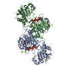
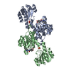
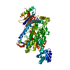
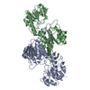
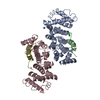
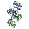
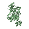
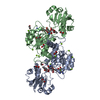
Mass: 33367.992 Da / Num. of mol.: 4
Source method: isolated from a genetically manipulated source
Source: (gene. exp.) Haloferax mediterranei ATCC 33500 (archaea)
Gene: ddh, serA5, HFX_2024 / Production host: Escherichia coli BL21(DE3) (bacteria)
References: UniProt: Q2VEQ7, Oxidoreductases; Acting on the CH-OH group of donors; With NAD+ or NADP+ as acceptor

-
Non-polymers , 7 types, 1798 molecules

#2: Chemical...
ChemComp-EDO / 1,2-ETHANEDIOL / ETHYLENE GLYCOL


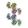
Mass: 62.068 Da / Num. of mol.: 22 / Source method: obtained synthetically / Formula: C2H6O2
#3: Chemical
ChemComp-SO4 / SULFATE ION


Mass: 96.063 Da / Num. of mol.: 4 / Source method: obtained synthetically / Formula: SO4
#4: Chemical...
ChemComp-MG / MAGNESIUM ION


Mass: 24.305 Da / Num. of mol.: 24 / Source method: obtained synthetically / Formula: Mg
#5: Chemical
ChemComp-NAD / NICOTINAMIDE-ADENINE-DINUCLEOTIDE


Mass: 663.425 Da / Num. of mol.: 4 / Source method: obtained synthetically / Formula: C21H27N7O14P2 / Comment: NAD*YM
#6: Chemical
ChemComp-7N5 / 2-Ketohexanoic acid


Mass: 130.142 Da / Num. of mol.: 4 / Source method: obtained synthetically / Formula: C6H10O3
#7: Chemical ChemComp-NA / SODIUM ION


Mass: 22.990 Da / Num. of mol.: 2 / Source method: obtained synthetically / Formula: Na
#8: Water ChemComp-HOH / water


Mass: 18.015 Da / Num. of mol.: 1738 / Source method: isolated from a natural source / Formula: H2O

-
Experimental details

-
Experiment

ExperimentMethod: X-RAY DIFFRACTION / Number of used crystals: 1

-
Sample preparation

CrystalDensity Matthews: 2.54 Å3/Da / Density % sol: 51.63 %
Crystal growTemperature: 290 K / Method: vapor diffusion, hanging drop / pH: 8
Details: Protein buffer: 20 mM Tris-HCl pH8, 3 mM EDTA and 1 M NaCl Crystallisation conditions: 0.1M Tris-HCl pH8, 0.5 M magnesium acetate and 20 % PEG3350 Ligands: 5 mM NAD+ and 50 mM 2-ketohexanoic acid

-
Data collection

DiffractionMean temperature: 100 K
Diffraction sourceSource: SYNCHROTRON / Site: Diamond / Beamline: I02 / Wavelength: 0.97204 Å
DetectorType: ADSC QUANTUM 315 / Detector: CCD / Date: Oct 26, 2008
RadiationProtocol: SINGLE WAVELENGTH / Monochromatic (M) / Laue (L): M / Scattering type: x-ray
Radiation wavelengthWavelength: 0.97204 Å / Relative weight: 1
ReflectionResolution: 1.35→46.54 Å / Num. obs: 268611 / % possible obs: 93 % / Redundancy: 2.1 % / Rmerge(I) obs: 0.061 / Net I/σ(I): 23.6
Reflection shellResolution: 1.35→1.37 Å / Redundancy: 2.2 % / Rmerge(I) obs: 0.462 / Mean I/σ(I) obs: 2.1 / % possible all: 91.7

-
Processing

Software
NameVersionClassification
REFMAC5.8.0135refinement
MOSFLMdata reduction
SCALAdata scaling
PHASERphasing
RefinementMethod to determine structure: MOLECULAR REPLACEMENT / Resolution: 1.35→46.54 Å / Cor.coef. Fo:Fc: 0.979 / Cor.coef. Fo:Fc free: 0.969 / SU B: 1.502 / SU ML: 0.027 / Cross valid method: THROUGHOUT / ESU R: 0.059 / ESU R Free: 0.055 / Details: HYDROGENS HAVE BEEN ADDED IN THE RIDING POSITIONS
RfactorNum. reflection% reflectionSelection details
Rfree0.16174 11807 5.1 %RANDOM
Rwork0.1264 ---
obs0.12817 219914 80.19 %-
Solvent computationIon probe radii: 0.8 Å / Shrinkage radii: 0.8 Å / VDW probe radii: 1.2 Å
Displacement parametersBiso mean: 17.066 Å2
Baniso -1Baniso -2Baniso -3
1-0.19 Å2-0.1 Å2-0.1 Å2
2--0.18 Å20.07 Å2
3----0.21 Å2
Refinement stepCycle: 1 / Resolution: 1.35→46.54 Å
ProteinNucleic acidLigandSolventTotal
Num. atoms9360 0 346 1738 11444
Refine LS restraints
Refine-IDTypeDev idealDev ideal targetNumber
X-RAY DIFFRACTIONr_bond_refined_d0.010.01910044
X-RAY DIFFRACTIONr_bond_other_d0.0020.029212
X-RAY DIFFRACTIONr_angle_refined_deg1.5021.98313729
X-RAY DIFFRACTIONr_angle_other_deg1.423321184
X-RAY DIFFRACTIONr_dihedral_angle_1_deg5.8951275
X-RAY DIFFRACTIONr_dihedral_angle_2_deg30.95223.693482
X-RAY DIFFRACTIONr_dihedral_angle_3_deg10.546151452
X-RAY DIFFRACTIONr_dihedral_angle_4_deg17.3151587
X-RAY DIFFRACTIONr_chiral_restr0.0830.21550
X-RAY DIFFRACTIONr_gen_planes_refined0.0080.02111468
X-RAY DIFFRACTIONr_gen_planes_other0.0030.022255
X-RAY DIFFRACTIONr_nbd_refined
X-RAY DIFFRACTIONr_nbd_other
X-RAY DIFFRACTIONr_nbtor_refined
X-RAY DIFFRACTIONr_nbtor_other
X-RAY DIFFRACTIONr_xyhbond_nbd_refined
X-RAY DIFFRACTIONr_xyhbond_nbd_other
X-RAY DIFFRACTIONr_metal_ion_refined
X-RAY DIFFRACTIONr_metal_ion_other
X-RAY DIFFRACTIONr_symmetry_vdw_refined
X-RAY DIFFRACTIONr_symmetry_vdw_other
X-RAY DIFFRACTIONr_symmetry_hbond_refined
X-RAY DIFFRACTIONr_symmetry_hbond_other
X-RAY DIFFRACTIONr_symmetry_metal_ion_refined
X-RAY DIFFRACTIONr_symmetry_metal_ion_other
X-RAY DIFFRACTIONr_mcbond_it2.8241.1974963
X-RAY DIFFRACTIONr_mcbond_other2.8251.1974962
X-RAY DIFFRACTIONr_mcangle_it3.0641.7946211
X-RAY DIFFRACTIONr_mcangle_other3.0651.7946212
X-RAY DIFFRACTIONr_scbond_it6.9461.6585081
X-RAY DIFFRACTIONr_scbond_other6.9551.6585065
X-RAY DIFFRACTIONr_scangle_it
X-RAY DIFFRACTIONr_scangle_other5.7922.2777470
X-RAY DIFFRACTIONr_long_range_B_refined5.65513.08613282
X-RAY DIFFRACTIONr_long_range_B_other5.16111.57112001
X-RAY DIFFRACTIONr_rigid_bond_restr14.562319256
X-RAY DIFFRACTIONr_sphericity_free27.8875408
X-RAY DIFFRACTIONr_sphericity_bonded21.444520428
LS refinement shellResolution: 1.35→1.385 Å / Total num. of bins used: 20
RfactorNum. reflection% reflection
Rfree0.254 441 -
Rwork0.221 8606 -
obs--42.16 %

+
About Yorodumi

-
News

-
Feb 9, 2022. New format data for meta-information of EMDB entries

New format data for meta-information of EMDB entries

  • Version 3 of the EMDB header file is now the official format.
  • The previous official version 1.9 will be removed from the archive.

Related info.:EMDB header

External links:wwPDB to switch to version 3 of the EMDB data model

-
Aug 12, 2020. Covid-19 info

Covid-19 info

URL: https://pdbjlvh1.pdbj.org/emnavi/covid19.php

New page: Covid-19 featured information page in EM Navigator.

Related info.:Covid-19 info / Mar 5, 2020. Novel coronavirus structure data

+
Mar 5, 2020. Novel coronavirus structure data

Novel coronavirus structure data

Related info.:Yorodumi Speices / Aug 12, 2020. Covid-19 info

External links:COVID-19 featured content - PDBj / Molecule of the Month (242):Coronavirus Proteases

+
Jan 31, 2019. EMDB accession codes are about to change! (news from PDBe EMDB page)

EMDB accession codes are about to change! (news from PDBe EMDB page)

  • The allocation of 4 digits for EMDB accession codes will soon come to an end. Whilst these codes will remain in use, new EMDB accession codes will include an additional digit and will expand incrementally as the available range of codes is exhausted. The current 4-digit format prefixed with “EMD-” (i.e. EMD-XXXX) will advance to a 5-digit format (i.e. EMD-XXXXX), and so on. It is currently estimated that the 4-digit codes will be depleted around Spring 2019, at which point the 5-digit format will come into force.
  • The EM Navigator/Yorodumi systems omit the EMD- prefix.

Related info.:Q: What is EMD? / ID/Accession-code notation in Yorodumi/EM Navigator

External links:EMDB Accession Codes are Changing Soon! / Contact to PDBj

+
Jul 12, 2017. Major update of PDB

Major update of PDB

  • wwPDB released updated PDB data conforming to the new PDBx/mmCIF dictionary.
  • This is a major update changing the version number from 4 to 5, and with Remediation, in which all the entries are updated.
  • In this update, many items about electron microscopy experimental information are reorganized (e.g. em_software).
  • Now, EM Navigator and Yorodumi are based on the updated data.

External links:wwPDB Remediation / Enriched Model Files Conforming to OneDep Data Standards Now Available in the PDB FTP Archive

-
Yorodumi

Thousand views of thousand structures

  • Yorodumi is a browser for structure data from EMDB, PDB, SASBDB, etc.
  • This page is also the successor to EM Navigator detail page, and also detail information page/front-end page for Omokage search.
  • The word "yorodu" (or yorozu) is an old Japanese word meaning "ten thousand". "mi" (miru) is to see.

Related info.:EMDB / PDB / SASBDB / Comparison of 3 databanks / Yorodumi Search / Aug 31, 2016. New EM Navigator & Yorodumi / Yorodumi Papers / Jmol/JSmol / Function and homology information / Changes in new EM Navigator and Yorodumi

Read more