| Entry | Database: PDB / ID: 6brk
|
|---|
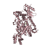
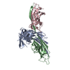
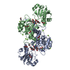
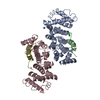
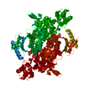
| Title | The SAM domain of mouse SAMHD1 is critical for its activation and regulation |
|---|
 Components Components | Deoxynucleoside triphosphate triphosphohydrolase SAMHD1 |
|---|
 Keywords Keywords | HYDROLASE / dNTPase / Allosteric Regulation / Binding Sites / Mouse / Models / Molecular / Protein Conformation / Protein Multimerization |
|---|
| Function / homology |  Function and homology information Function and homology information
Nucleotide catabolism / triphosphoric monoester hydrolase activity / Hydrolases; Acting on ester bonds; Triphosphoric-monoester hydrolases / deoxynucleoside triphosphate hydrolase activity / dGTP binding / dATP catabolic process / deoxyribonucleotide catabolic process / tetraspanin-enriched microdomain / dGTPase activity / dGTP catabolic process ...Nucleotide catabolism / triphosphoric monoester hydrolase activity / Hydrolases; Acting on ester bonds; Triphosphoric-monoester hydrolases / deoxynucleoside triphosphate hydrolase activity / dGTP binding / dATP catabolic process / deoxyribonucleotide catabolic process / tetraspanin-enriched microdomain / dGTPase activity / dGTP catabolic process / DNA strand resection involved in replication fork processing / negative regulation of type I interferon-mediated signaling pathway / regulation of innate immune response / RNA nuclease activity / somatic hypermutation of immunoglobulin genes / double-strand break repair via homologous recombination / single-stranded DNA binding / site of double-strand break / defense response to virus / protein homotetramerization / nucleic acid binding / innate immune response / DNA damage response / GTP binding / RNA binding / zinc ion binding / nucleoplasm / identical protein binding / nucleus / plasma membraneSimilarity search - Function : / HD domain profile. / HD domain / HD domain / SAM domain (Sterile alpha motif) / SAM domain profile. / Metal dependent phosphohydrolases with conserved 'HD' motif. / HD/PDEase domain / Sterile alpha motif. / Sterile alpha motif domain / Sterile alpha motif/pointed domain superfamilySimilarity search - Domain/homology 2'-DEOXYGUANOSINE-5'-TRIPHOSPHATE / Deoxynucleoside triphosphate triphosphohydrolase SAMHD1 / Deoxynucleoside triphosphate triphosphohydrolase SAMHD1Similarity search - Component |
|---|
| Biological species |   Mus musculus (house mouse) Mus musculus (house mouse) |
|---|
| Method |  X-RAY DIFFRACTION / SYNCHROTRON / MOLECULAR REPLACEMENT / Resolution: 3.5 Å X-RAY DIFFRACTION / SYNCHROTRON / MOLECULAR REPLACEMENT / Resolution: 3.5 Å |
|---|
 Authors Authors | Buzovetsky, O. / Tang, C. / Knecht, K.M. / Antonucci, J.M. / Wu, L. / Ji, X. / Xiong, Y. |
|---|
 Citation Citation | Journal: Nat Commun / Year: 2018
Title: The SAM domain of mouse SAMHD1 is critical for its activation and regulation.
Authors: Buzovetsky, O. / Tang, C. / Knecht, K.M. / Antonucci, J.M. / Wu, L. / Ji, X. / Xiong, Y. |
|---|
| History | | Deposition | Nov 30, 2017 | Deposition site: RCSB / Processing site: RCSB |
|---|
| Revision 1.0 | Feb 14, 2018 | Provider: repository / Type: Initial release |
|---|
| Revision 1.1 | Oct 4, 2023 | Group: Data collection / Database references / Refinement description
Category: chem_comp_atom / chem_comp_bond ...chem_comp_atom / chem_comp_bond / database_2 / pdbx_initial_refinement_model
Item: _database_2.pdbx_DOI / _database_2.pdbx_database_accession |
|---|
| Revision 1.2 | Nov 6, 2024 | Group: Structure summary / Category: pdbx_entry_details / pdbx_modification_feature |
|---|
|
|---|
 Movie
Movie Controller

 Yorodumi
 Open data
 Basic information
 Structure visualization
 Downloads & links
 Other downloads


 PDBj
PDBj
 Assembly
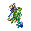

 Mass: 507.181 Da / Num. of mol.: 3 / Source method: obtained synthetically / Formula: C10H16N5O13P3
 Mass: 24.305 Da / Num. of mol.: 1 / Source method: obtained synthetically / Formula: Mg
 Sample preparation
 / Beamline: 24-ID-C / Wavelength: 0.9164 Å
 Processing