 Movie
Movie Controller


+ Open data

- Basic information
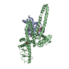
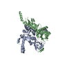
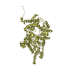
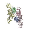
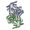
| Entry | Database: PDB / ID: 5lii | ||||||
|---|---|---|---|---|---|---|---|
| Title | bacteriophage phi812K1-420 major capsid protein | ||||||
 Components Components | Major capsid protein | ||||||
 Keywords Keywords | VIRUS LIKE PARTICLE / polyvalent staphylococcal bactoriophage / Myoviridae / tail sheath / tail contraction | ||||||
| Function / homology | Major capsid protein Function and homology information Function and homology information | ||||||
| Biological species |  Staphylococcus phage 812 (virus) Staphylococcus phage 812 (virus) | ||||||
| Method | ELECTRON MICROSCOPY / single particle reconstruction / cryo EM / Resolution: 3.8 Å | ||||||
 Authors Authors | Novacek, J. / Siborova, M. / Benesik, M. / Pantucek, R. / Doskar, J. / Plevka, P. | ||||||
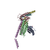
 Citation Citation | Journal: Proc Natl Acad Sci U S A / Year: 2016 Title: Structure and genome release of Twort-like Myoviridae phage with a double-layered baseplate. Authors: Jiří Nováček / Marta Šiborová / Martin Benešík / Roman Pantůček / Jiří Doškař / Pavel Plevka /  Abstract: Bacteriophages from the family Myoviridae use double-layered contractile tails to infect bacteria. Contraction of the tail sheath enables the tail tube to penetrate through the bacterial cell wall ...Bacteriophages from the family Myoviridae use double-layered contractile tails to infect bacteria. Contraction of the tail sheath enables the tail tube to penetrate through the bacterial cell wall and serve as a channel for the transport of the phage genome into the cytoplasm. However, the mechanisms controlling the tail contraction and genome release of phages with "double-layered" baseplates were unknown. We used cryo-electron microscopy to show that the binding of the Twort-like phage phi812 to the Staphylococcus aureus cell wall requires a 210° rotation of the heterohexameric receptor-binding and tripod protein complexes within its baseplate about an axis perpendicular to the sixfold axis of the tail. This rotation reorients the receptor-binding proteins to point away from the phage head, and also results in disruption of the interaction of the tripod proteins with the tail sheath, hence triggering its contraction. However, the tail sheath contraction of Myoviridae phages is not sufficient to induce genome ejection. We show that the end of the phi812 double-stranded DNA genome is bound to one protein subunit from a connector complex that also forms an interface between the phage head and tail. The tail sheath contraction induces conformational changes of the neck and connector that result in disruption of the DNA binding. The genome penetrates into the neck, but is stopped at a bottleneck before the tail tube. A subsequent structural change of the tail tube induced by its interaction with the S. aureus cell is required for the genome's release. | ||||||
| History |
|
- Structure visualization
| Movie |
Movie viewer |
|---|---|
| Structure viewer | Molecule: MolmilJmol/JSmol |
- Downloads & links
-Download
| PDBx/mmCIF format | 5lii.cif.gz | 90.8 KB | Display | PDBx/mmCIF format |
|---|---|---|---|---|
| PDB format | pdb5lii.ent.gz | 66.7 KB | Display | PDB format |
| PDBx/mmJSON format | 5lii.json.gz | Tree view | PDBx/mmJSON format | |
| Others |  Other downloads Other downloads |
-Validation report
| Arichive directory | https://data.pdbj.org/pub/pdb/validation_reports/li/5liiftp://data.pdbj.org/pub/pdb/validation_reports/li/5lii | HTTPS FTP |
|---|
-Related structure data
| Related structure data |  4053MC 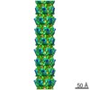 4003C  4051C  4052C 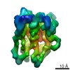 4054C  8201C  8202C  8203C  8204C 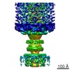 8205C  8206C 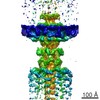 8207C  8208C  8209C 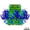 8210C  8211C 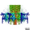 8212C  8213C  8214C  8304C  5li2C  5li4C  5lijC M: map data used to model this data C: citing same article ( |
|---|---|
| Similar structure data |

-Links
 PDBj
PDBj- Assembly
| Deposited unit | 
|
|---|---|
| 1 |
|
-Components
| #1: Protein | Mass: 51288.707 Da / Num. of mol.: 1 Source method: isolated from a genetically manipulated source Source: (gene. exp.) Staphylococcus phage 812 (virus) / Gene: 812_096, 812a_096, 812F1_096, K1/420_096, K1_096 / Production host:  Staphylococcaceae (Staphylococcus group) / References: UniProt: A1YTN4 Staphylococcaceae (Staphylococcus group) / References: UniProt: A1YTN4 |
|---|
-Experimental details
-Experiment
| Experiment | Method: ELECTRON MICROSCOPY |
|---|---|
| EM experiment | Aggregation state: PARTICLE / 3D reconstruction method: single particle reconstruction |
- Sample preparation
| Component | Name: Staphylococcus phage 812 / Type: VIRUS / Entity ID: all / Source: NATURAL | ||||||||||||||||||||
|---|---|---|---|---|---|---|---|---|---|---|---|---|---|---|---|---|---|---|---|---|---|
| Molecular weight | Experimental value: NO | ||||||||||||||||||||
| Source (natural) | Organism: Staphylococcus phage 812 (virus) / Strain: K420 | ||||||||||||||||||||
| Details of virus | Empty: NO / Enveloped: NO / Isolate: SPECIES / Type: VIRION | ||||||||||||||||||||
| Natural host | Organism: Staphylococcus aureus | ||||||||||||||||||||
| Virus shell | Diameter: 1100 nm / Triangulation number (T number): 16 | ||||||||||||||||||||
| Buffer solution | pH: 8 | ||||||||||||||||||||
| Buffer component |
| ||||||||||||||||||||
| Specimen | Conc.: 1 mg/ml / Embedding applied: NO / Shadowing applied: NO / Staining applied: NO / Vitrification applied: YES | ||||||||||||||||||||
| Specimen support | Grid material: COPPER / Grid mesh size: 200 divisions/in. / Grid type: Quantifoil R2/1 | ||||||||||||||||||||
| Vitrification | Instrument: FEI VITROBOT MARK IV / Cryogen name: ETHANE / Humidity: 100 % / Chamber temperature: 295 K |
- Electron microscopy imaging
| Experimental equipment |  Model: Titan Krios / Image courtesy: FEI Company |
|---|---|
| Microscopy | Model: FEI TITAN KRIOS |
| Electron gun | Electron source:  FIELD EMISSION GUN / Accelerating voltage: 300 kV / Illumination mode: FLOOD BEAM FIELD EMISSION GUN / Accelerating voltage: 300 kV / Illumination mode: FLOOD BEAM |
| Electron lens | Mode: BRIGHT FIELD / Cs: 2.7 mm / C2 aperture diameter: 100 µm / Alignment procedure: COMA FREE |
| Specimen holder | Cryogen: NITROGEN / Specimen holder model: FEI TITAN KRIOS AUTOGRID HOLDER |
| Image recording | Average exposure time: 0.86 sec. / Electron dose: 20 e/Å2 / Detector mode: INTEGRATING / Film or detector model: FEI FALCON II (4k x 4k) |
| Image scans | Movie frames/image: 7 |
- Processing
| EM software |
| ||||||||||||||||||||||||||||
|---|---|---|---|---|---|---|---|---|---|---|---|---|---|---|---|---|---|---|---|---|---|---|---|---|---|---|---|---|---|
| CTF correction | Type: PHASE FLIPPING ONLY | ||||||||||||||||||||||||||||
| Symmetry | Point symmetry: I (icosahedral) | ||||||||||||||||||||||||||||
| 3D reconstruction | Resolution: 3.8 Å / Resolution method: FSC 0.143 CUT-OFF / Num. of particles: 12018 / Algorithm: FOURIER SPACE / Symmetry type: HELICAL |
