 Movie
Movie Controller

[English] 日本語
 Yorodumi
- PDB-5la2: The mechanism by which arabinoxylanases can recognise highly deco... -
+ Open data

- Basic information
| Entry | Database: PDB / ID: 5la2 | |||||||||
|---|---|---|---|---|---|---|---|---|---|---|
| Title | The mechanism by which arabinoxylanases can recognise highly decorated xylans | |||||||||
 Components Components | Carbohydrate binding family 6 | |||||||||
 Keywords Keywords | HYDROLASE / Arabinoxylanase / Glycoside hydrolase / Carbohydrate binding module / Arabinose / Clostridium thermocellum / Cellulosome | |||||||||
| Function / homology |  Function and homology information Function and homology informationhydrolase activity, hydrolyzing O-glycosyl compounds / cellulose catabolic process / carbohydrate binding / metal ion binding Similarity search - Function | |||||||||
| Biological species |  Ruminiclostridium thermocellum (bacteria) Ruminiclostridium thermocellum (bacteria) | |||||||||
| Method |  X-RAY DIFFRACTION / SYNCHROTRON / MOLECULAR REPLACEMENT / Resolution: 1.65 Å X-RAY DIFFRACTION / SYNCHROTRON / MOLECULAR REPLACEMENT / Resolution: 1.65 Å | |||||||||
 Authors Authors | Basle, A. / Labourel, A. / Cuskin, F. / Jackson, A. / Crouch, L. / Rogowski, A. / Gilbert, H. | |||||||||
| Funding support |  United Kingdom, 1items United Kingdom, 1items
| |||||||||
 Citation Citation | Journal: J.Biol.Chem. / Year: 2016 Title: The Mechanism by Which Arabinoxylanases Can Recognize Highly Decorated Xylans. Authors: Labourel, A. / Crouch, L.I. / Bras, J.L. / Jackson, A. / Rogowski, A. / Gray, J. / Yadav, M.P. / Henrissat, B. / Fontes, C.M. / Gilbert, H.J. / Najmudin, S. / Basle, A. / Cuskin, F. | |||||||||
| History |
|
- Structure visualization
| Structure viewer | Molecule: MolmilJmol/JSmol |
|---|
- Downloads & links
-Download
| PDBx/mmCIF format | 5la2.cif.gz | 420.3 KB | Display | PDBx/mmCIF format |
|---|---|---|---|---|
| PDB format | pdb5la2.ent.gz | 338.6 KB | Display | PDB format |
| PDBx/mmJSON format | 5la2.json.gz | Tree view | PDBx/mmJSON format | |
| Others |  Other downloads Other downloads |
-Validation report
| Arichive directory | https://data.pdbj.org/pub/pdb/validation_reports/la/5la2ftp://data.pdbj.org/pub/pdb/validation_reports/la/5la2 | HTTPS FTP |
|---|
-Related structure data
| Related structure data |  5g56C  5la0C  5la1C  5ak1 S: Starting model for refinement C: citing same article ( |
|---|---|
| Similar structure data |

-Links
 PDBj
PDBj

- Assembly
| Deposited unit | 
| ||||||||
|---|---|---|---|---|---|---|---|---|---|
| 1 | 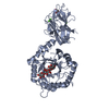
| ||||||||
| 2 | 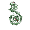
| ||||||||
| Unit cell |
|
-Components
| #1: Protein | Mass: 54115.652 Da / Num. of mol.: 2 / Fragment: UNP residues 37-516 Source method: isolated from a genetically manipulated source Source: (gene. exp.) Ruminiclostridium thermocellum (bacteria)Gene: Cther_1146 / Production host: #2: Polysaccharide | Source method: isolated from a genetically manipulated source #3: Sugar |   Type: L-saccharide, beta linking / Mass: 150.130 Da / Num. of mol.: 2 Type: L-saccharide, beta linking / Mass: 150.130 Da / Num. of mol.: 2Source method: isolated from a genetically manipulated source Formula: C5H10O5 #4: Chemical | ChemComp-CA /   Mass: 40.078 Da / Num. of mol.: 6 / Source method: obtained synthetically / Formula: Ca Mass: 40.078 Da / Num. of mol.: 6 / Source method: obtained synthetically / Formula: Ca#5: Water | ChemComp-HOH / |  Mass: 18.015 Da / Num. of mol.: 923 / Source method: isolated from a natural source / Formula: H2O Mass: 18.015 Da / Num. of mol.: 923 / Source method: isolated from a natural source / Formula: H2O |
|---|
-Experimental details
-Experiment
| Experiment | Method: X-RAY DIFFRACTION / Number of used crystals: 1 |
|---|
- Sample preparation
| Crystal | Density Matthews: 2.72 Å3/Da / Density % sol: 54.79 % |
|---|---|
| Crystal grow | Temperature: 293.15 K / Method: vapor diffusion Details: 100 mM Tris-Bicine buffer pH 8.5, 12.5% (w/v) polyethylene glycol average Mw 1,000 Da, 12.5% (w/v) polyethylene glycol average Mw 3,350 Da and 12.5% (RS)-2-methyl-2,4-pentanediol (racemic). |
-Data collection
| Diffraction | Mean temperature: 100 K |
|---|---|
| Diffraction source | Source: SYNCHROTRON / Site: Diamond / Beamline: I02 / Wavelength: 0.9791 Å |
| Detector | Type: DECTRIS PILATUS3 6M / Detector: PIXEL / Date: Sep 20, 2015 |
| Radiation | Protocol: SINGLE WAVELENGTH / Monochromatic (M) / Laue (L): M / Scattering type: x-ray |
| Radiation wavelength | Wavelength: 0.9791 Å / Relative weight: 1 |
| Reflection | Resolution: 1.65→48.43 Å / Num. obs: 140288 / % possible obs: 99.8 % / Observed criterion σ(I): 1.5 / Redundancy: 3.3 % / CC1/2: 0.988 / Rmerge(I) obs: 0.057 / Net I/σ(I): 11.2 |
| Reflection shell | Resolution: 1.65→1.68 Å / Redundancy: 3.4 % / Rmerge(I) obs: 0.749 / Mean I/σ(I) obs: 1.6 / % possible all: 99.4 |
- Processing
| Software |
| ||||||||||||||||||||||||||||||||||||||||||||||||||||||||||||||||||||||||||||||||||||||||||||||||||||||||||||||||||||||||||||||||||||||||||||||||||||||||||||||||||||||||||||||||||||||
|---|---|---|---|---|---|---|---|---|---|---|---|---|---|---|---|---|---|---|---|---|---|---|---|---|---|---|---|---|---|---|---|---|---|---|---|---|---|---|---|---|---|---|---|---|---|---|---|---|---|---|---|---|---|---|---|---|---|---|---|---|---|---|---|---|---|---|---|---|---|---|---|---|---|---|---|---|---|---|---|---|---|---|---|---|---|---|---|---|---|---|---|---|---|---|---|---|---|---|---|---|---|---|---|---|---|---|---|---|---|---|---|---|---|---|---|---|---|---|---|---|---|---|---|---|---|---|---|---|---|---|---|---|---|---|---|---|---|---|---|---|---|---|---|---|---|---|---|---|---|---|---|---|---|---|---|---|---|---|---|---|---|---|---|---|---|---|---|---|---|---|---|---|---|---|---|---|---|---|---|---|---|---|---|
| Refinement | Method to determine structure: MOLECULAR REPLACEMENT Starting model: 5AK1 5ak1 Resolution: 1.65→48.43 Å / Cor.coef. Fo:Fc: 0.972 / Cor.coef. Fo:Fc free: 0.953 / SU B: 4.545 / SU ML: 0.066 / Cross valid method: THROUGHOUT / ESU R: 0.094 / ESU R Free: 0.084 / Details: HYDROGENS HAVE BEEN ADDED IN THE RIDING POSITIONS
| ||||||||||||||||||||||||||||||||||||||||||||||||||||||||||||||||||||||||||||||||||||||||||||||||||||||||||||||||||||||||||||||||||||||||||||||||||||||||||||||||||||||||||||||||||||||
| Solvent computation | Ion probe radii: 0.8 Å / Shrinkage radii: 0.8 Å / VDW probe radii: 1.2 Å | ||||||||||||||||||||||||||||||||||||||||||||||||||||||||||||||||||||||||||||||||||||||||||||||||||||||||||||||||||||||||||||||||||||||||||||||||||||||||||||||||||||||||||||||||||||||
| Displacement parameters | Biso mean: 21.695 Å2
| ||||||||||||||||||||||||||||||||||||||||||||||||||||||||||||||||||||||||||||||||||||||||||||||||||||||||||||||||||||||||||||||||||||||||||||||||||||||||||||||||||||||||||||||||||||||
| Refinement step | Cycle: 1 / Resolution: 1.65→48.43 Å
| ||||||||||||||||||||||||||||||||||||||||||||||||||||||||||||||||||||||||||||||||||||||||||||||||||||||||||||||||||||||||||||||||||||||||||||||||||||||||||||||||||||||||||||||||||||||
| Refine LS restraints |
|