 Movie
Movie Controller

[English] 日本語
 Yorodumi
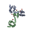
- PDB-5g56: THE TETRA-MODULAR CELLULOSOMAL ARABINOXYLANASE CtXyl5A STRUCTURE ... -
+ Open data

- Basic information
| Entry | Database: PDB / ID: 5g56 | |||||||||
|---|---|---|---|---|---|---|---|---|---|---|
| Title | THE TETRA-MODULAR CELLULOSOMAL ARABINOXYLANASE CtXyl5A STRUCTURE AS REVEALED BY X-RAY CRYSTALLOGRAPHY | |||||||||
 Components Components | CARBOHYDRATE BINDING FAMILY 6 | |||||||||
 Keywords Keywords | CARBOHYDRATE BINDING PROTEIN / ARABINOXYLANASE / CTXYL5A / GH5 / CBM6 / CBM13 / FN3 / CLOSTRIDIUM THERMOCELLUM / CELLULOSOME | |||||||||
| Function / homology |  Function and homology information Function and homology informationhydrolase activity, hydrolyzing O-glycosyl compounds / cellulose catabolic process / carbohydrate binding / metal ion binding Similarity search - Function | |||||||||
| Biological species |  CLOSTRIDIUM THERMOCELLUM (bacteria) CLOSTRIDIUM THERMOCELLUM (bacteria) | |||||||||
| Method |  X-RAY DIFFRACTION / SYNCHROTRON / MOLECULAR REPLACEMENT / Resolution: 2.64 Å X-RAY DIFFRACTION / SYNCHROTRON / MOLECULAR REPLACEMENT / Resolution: 2.64 Å | |||||||||
 Authors Authors | Bras, J.L.A. / Gilbert, H.J. / Ferreira, L.M.A. / Fontes, C.M.G.A. / Najmudin, S. | |||||||||
 Citation Citation | Journal: J.Biol.Chem. / Year: 2016 Title: The Mechanism by which Arabinoxylanases Can Recognise Highly Decorated Xylans. Authors: Labourel, A. / Crouch, L.I. / Bras, J.L. / Jackson, A. / Rogowski, A. / Gray, J. / Yadav, M.P. / Henrissat, B. / Fontes, C.M. / Gilbert, H.J. / Najmudin, S. / Basle, A. / Cuskin, F. #1: Journal: Acta Crystallogr.,Sect.F / Year: 2011 Title: Purification, Crystallization and Preliminary X-Ray Characterization of the Pentamodular Arabinoxylanase Ctxyl5A from Clostridium Thermocellum. Authors: Bras, J.L.A. / Correia, M.A.S. / Romao, M.J. / Prates, J.A.M. / Fontes, C.M.G.A. / Najmudin, S. | |||||||||
| History |
| |||||||||
| Remark 700 | SHEET DETERMINATION METHOD: DSSP THE SHEETS PRESENTED AS "AL" IN EACH CHAIN ON SHEET RECORDS BELOW ... SHEET DETERMINATION METHOD: DSSP THE SHEETS PRESENTED AS "AL" IN EACH CHAIN ON SHEET RECORDS BELOW IS ACTUALLY AN 6-STRANDED BARREL THIS IS REPRESENTED BY A 7-STRANDED SHEET IN WHICH THE FIRST AND LAST STRANDS ARE IDENTICAL. |
- Structure visualization
| Structure viewer | Molecule: MolmilJmol/JSmol |
|---|
- Downloads & links
-Download
| PDBx/mmCIF format | 5g56.cif.gz | 290.8 KB | Display | PDBx/mmCIF format |
|---|---|---|---|---|
| PDB format | pdb5g56.ent.gz | 232.6 KB | Display | PDB format |
| PDBx/mmJSON format | 5g56.json.gz | Tree view | PDBx/mmJSON format | |
| Others |  Other downloads Other downloads |
-Validation report
| Arichive directory | https://data.pdbj.org/pub/pdb/validation_reports/g5/5g56ftp://data.pdbj.org/pub/pdb/validation_reports/g5/5g56 | HTTPS FTP |
|---|
-Related structure data
| Related structure data |  5la0C 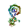 5la1C  5la2C  2y8kS  2y8m 2y9i 2y9s  3mpcS S: Starting model for refinement C: citing same article ( |
|---|---|
| Similar structure data |


-Links
 PDBj
PDBj

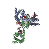
- Assembly
| Deposited unit | 
| ||||||||
|---|---|---|---|---|---|---|---|---|---|
| 1 |
| ||||||||
| Unit cell |
|
-Components
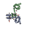
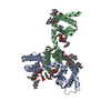
| #1: Protein | Mass: 93949.648 Da / Num. of mol.: 1 / Fragment: RESIDUES 36-889 Source method: isolated from a genetically manipulated source Details: SEMET DERIVATIVE / Source: (gene. exp.) CLOSTRIDIUM THERMOCELLUM (bacteria) / Production host: | ||||||||
|---|---|---|---|---|---|---|---|---|---|
| #2: Chemical |   Mass: 40.078 Da / Num. of mol.: 3 / Source method: obtained synthetically / Formula: Ca Mass: 40.078 Da / Num. of mol.: 3 / Source method: obtained synthetically / Formula: Ca#3: Chemical |   Mass: 118.174 Da / Num. of mol.: 2 / Source method: obtained synthetically / Formula: C6H14O2 / Comment: precipitant*YM Mass: 118.174 Da / Num. of mol.: 2 / Source method: obtained synthetically / Formula: C6H14O2 / Comment: precipitant*YM#4: Water | ChemComp-HOH / |  Mass: 18.015 Da / Num. of mol.: 227 / Source method: isolated from a natural source / Formula: H2O Mass: 18.015 Da / Num. of mol.: 227 / Source method: isolated from a natural source / Formula: H2OHas protein modification | Y | Sequence details | SEMET DERIVATIVE | |
-Experimental details
-Experiment
| Experiment | Method: X-RAY DIFFRACTION / Number of used crystals: 1 |
|---|
- Sample preparation
| Crystal | Density Matthews: 3.66 Å3/Da / Density % sol: 68 % / Description: CBM13 MODEL WAS BUILT IN MANUALLY USING COOT. |
|---|---|
| Crystal grow | pH: 7 / Details: 40% (V/V) 2-METHYL-2-4PENTANEDIOL (MPD), pH 7 |
-Data collection
| Diffraction | Mean temperature: 100 K |
|---|---|
| Diffraction source | Source: SYNCHROTRON / Site: ESRF  / Beamline: ID14-4 / Wavelength: 0.9334 / Beamline: ID14-4 / Wavelength: 0.9334 |
| Detector | Type: ADSC QUANTUM 315 / Detector: CCD / Date: Dec 4, 2009 |
| Radiation | Protocol: SINGLE WAVELENGTH / Monochromatic (M) / Laue (L): M / Scattering type: x-ray |
| Radiation wavelength | Wavelength: 0.9334 Å / Relative weight: 1 |
| Reflection | Resolution: 2.64→50.7 Å / Num. obs: 42246 / % possible obs: 98.5 % / Observed criterion σ(I): 0 / Redundancy: 5.8 % / Rmerge(I) obs: 0.16 / Net I/σ(I): 8 |
| Reflection shell | Resolution: 2.64→2.78 Å / Redundancy: 5 % / Rmerge(I) obs: 0.7 / Mean I/σ(I) obs: 2 / % possible all: 96.4 |
- Processing
| Software |
| ||||||||||||||||||||||||||||||||||||||||||||||||||||||||||||||||||||||||||||||||||||||||||||||||||||||||||||||||||||||||||||||||||||||||||||||||||||||||||||||||||||||||||||||||||||||
|---|---|---|---|---|---|---|---|---|---|---|---|---|---|---|---|---|---|---|---|---|---|---|---|---|---|---|---|---|---|---|---|---|---|---|---|---|---|---|---|---|---|---|---|---|---|---|---|---|---|---|---|---|---|---|---|---|---|---|---|---|---|---|---|---|---|---|---|---|---|---|---|---|---|---|---|---|---|---|---|---|---|---|---|---|---|---|---|---|---|---|---|---|---|---|---|---|---|---|---|---|---|---|---|---|---|---|---|---|---|---|---|---|---|---|---|---|---|---|---|---|---|---|---|---|---|---|---|---|---|---|---|---|---|---|---|---|---|---|---|---|---|---|---|---|---|---|---|---|---|---|---|---|---|---|---|---|---|---|---|---|---|---|---|---|---|---|---|---|---|---|---|---|---|---|---|---|---|---|---|---|---|---|---|
| Refinement | Method to determine structure: MOLECULAR REPLACEMENT Starting model: PDB ENTRIES 2Y8K, 3MPC, 2Y8M, 2Y9I AND 2Y9S Resolution: 2.64→50.7 Å / Cor.coef. Fo:Fc: 0.892 / Cor.coef. Fo:Fc free: 0.85 / SU B: 22.967 / SU ML: 0.245 / Cross valid method: THROUGHOUT / ESU R: 0.369 / ESU R Free: 0.284 / Stereochemistry target values: MAXIMUM LIKELIHOOD Details: HYDROGENS HAVE BEEN ADDED IN THE RIDING POSITIONS. U VALUES WITH TLS ADDED FOR GH5, CBM6,CBM13 AND FN3 MODULES. CBM62 WAS NOT FITTED IN THE SOLVENT CHANNEL. DISORDERED REGIONS WERE MODELED STEREOCHEMICALLY
| ||||||||||||||||||||||||||||||||||||||||||||||||||||||||||||||||||||||||||||||||||||||||||||||||||||||||||||||||||||||||||||||||||||||||||||||||||||||||||||||||||||||||||||||||||||||
| Solvent computation | Ion probe radii: 1 Å / Shrinkage radii: 1 Å / VDW probe radii: 1.4 Å / Solvent model: MASK | ||||||||||||||||||||||||||||||||||||||||||||||||||||||||||||||||||||||||||||||||||||||||||||||||||||||||||||||||||||||||||||||||||||||||||||||||||||||||||||||||||||||||||||||||||||||
| Refinement step | Cycle: LAST / Resolution: 2.64→50.7 Å
| ||||||||||||||||||||||||||||||||||||||||||||||||||||||||||||||||||||||||||||||||||||||||||||||||||||||||||||||||||||||||||||||||||||||||||||||||||||||||||||||||||||||||||||||||||||||
| Refine LS restraints |
|