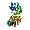
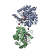
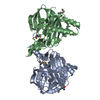
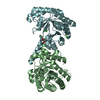
- PDB-2y8k: Structure of CtGH5-CBM6, an arabinoxylan-specific xylanase. -
+
Open data
ID or keywords:
Loading...
-
Basic information
Entry
Database: PDB / ID: 2y8k
Title
Structure of CtGH5-CBM6, an arabinoxylan-specific xylanase.
Components
CARBOHYDRATE BINDING FAMILY 6
Keywords
HYDROLASE
Function / homology
Function and homology information
hydrolase activity, hydrolyzing O-glycosyl compounds / cellulose catabolic process / carbohydrate binding / metal ion binding Similarity search - Function
Cellulose binding, type IV / Cellulose Binding Domain Type IV / Ricin-type beta-trefoil lectin domain-like / Carbohydrate binding module (family 6) / CBM6 (carbohydrate binding type-6) domain profile. / Carbohydrate binding module family 6 / Glycoside hydrolase, family 5, conserved site / Glycosyl hydrolases family 5 signature. / Dockerin domain / Dockerin domain profile. ...Cellulose binding, type IV / Cellulose Binding Domain Type IV / Ricin-type beta-trefoil lectin domain-like / Carbohydrate binding module (family 6) / CBM6 (carbohydrate binding type-6) domain profile. / Carbohydrate binding module family 6 / Glycoside hydrolase, family 5, conserved site / Glycosyl hydrolases family 5 signature. / Dockerin domain / Dockerin domain profile. / Dockerin type I domain / Dockerin type I repeat / Dockerin domain superfamily / Cellulase (glycosyl hydrolase family 5) / Glycoside hydrolase, family 5 / Ricin B, lectin domain / Ricin B-like lectins / Galactose-binding domain-like / Fibronectin type-III domain profile. / Galactose-binding-like domain superfamily / Fibronectin type III / Fibronectin type III superfamily / EF-Hand 1, calcium-binding site / EF-hand calcium-binding domain. / Glycosidases / Glycoside hydrolase superfamily / Jelly Rolls / TIM Barrel / Alpha-Beta Barrel / Immunoglobulin-like fold / Sandwich / Mainly Beta / Alpha Beta Similarity search - Domain/homology
SHEET DETERMINATION METHOD: DSSP THE SHEETS PRESENTED AS "AB" IN EACH CHAIN ON SHEET RECORDS BELOW ... SHEET DETERMINATION METHOD: DSSP THE SHEETS PRESENTED AS "AB" IN EACH CHAIN ON SHEET RECORDS BELOW IS ACTUALLY AN 8-STRANDED BARREL THIS IS REPRESENTED BY A 9-STRANDED SHEET IN WHICH THE FIRST AND LAST STRANDS ARE IDENTICAL.
Mass: 18.015 Da / Num. of mol.: 625 / Source method: isolated from a natural source / Formula: H2O
Sequence details
ONLY GH5 AND CBM6 MODULES WERE CLONED FOR CRYSTALLISATION
-
Experimental details
-
Experiment
Experiment
Method: X-RAY DIFFRACTION / Number of used crystals: 2
-
Sample preparation
Crystal
Density Matthews: 2.6 Å3/Da / Density % sol: 52 % Description: SOLVED BY SAD WITH ASSOCIATED DATASET AND REFINED TO THE HIGHER RESOLUTION OF THIS DATASET. SELENOMETHIONINE PROTEIN HAD 2 ENGINEERED METHIONINES IN THE SEQUENCE. NATIVE DATA IS FROM NATIVE SEQUENCE.
Resolution: 1.47→36.74 Å / Num. obs: 94925 / % possible obs: 99.9 % / Observed criterion σ(I): 0 / Redundancy: 7 % / Biso Wilson estimate: 16.2 Å2 / Rmerge(I) obs: 0.08 / Net I/σ(I): 13.9
Reflection shell
Resolution: 1.47→1.55 Å / Redundancy: 6.6 % / Rmerge(I) obs: 0.64 / Mean I/σ(I) obs: 2.8 / % possible all: 99.9
-
Processing
Software
Name
Version
Classification
REFMAC
5.5.0109
refinement
MOSFLM
datareduction
SCALA
datascaling
SHELXCDE
phasing
Refinement
Method to determine structure: SAD Starting model: NONE Resolution: 1.47→35.58 Å / Cor.coef. Fo:Fc: 0.976 / Cor.coef. Fo:Fc free: 0.97 / SU B: 0.903 / SU ML: 0.034 / Cross valid method: THROUGHOUT / ESU R: 0.054 / ESU R Free: 0.055 / Stereochemistry target values: MAXIMUM LIKELIHOOD / Details: HYDROGENS HAVE BEEN ADDED IN THE RIDING POSITIONS.
Rfactor
Num. reflection
% reflection
Selection details
Rfree
0.16627
4744
5 %
RANDOM
Rwork
0.14581
-
-
-
obs
0.14685
90100
99.95 %
-
Solvent computation
Ion probe radii: 0.8 Å / Shrinkage radii: 0.8 Å / VDW probe radii: 1.2 Å / Solvent model: BABINET MODEL WITH MASK
 Movie
Movie Controller


 Open data
 Basic information
 Components
 Keywords
 Function and homology information
 CLOSTRIDIUM THERMOCELLUM (bacteria)
 X-RAY DIFFRACTION /
  Authors
 Citation
 Structure visualization
 Downloads & links
 Other downloads
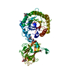

 PDBj
PDBj

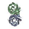
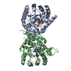
 Assembly
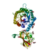

 Mass: 92.094 Da / Num. of mol.: 8 / Source method: obtained synthetically / Formula: C3H8O3
 Mass: 40.078 Da / Num. of mol.: 1 / Source method: obtained synthetically / Formula: Ca
 Mass: 22.990 Da / Num. of mol.: 4 / Source method: obtained synthetically / Formula: Na
 Mass: 18.015 Da / Num. of mol.: 625 / Source method: isolated from a natural source / Formula: H2O
 Sample preparation

 Processing