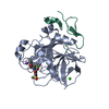
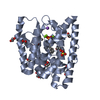
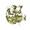
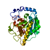
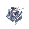
Entry Database : PDB / ID : 5j6dTitle Discovery of acyl guanidine tryptophan hydroxylase-1 inhibitors Tryptophan 5-hydroxylase 1 Keywords / / / / / Function / homology Function Domain/homology Component
/ / / / / / / / / / / / / / / / / / / / / / / / / / / / / / / / / / / / / / / / / / / / / / / / Biological species Homo sapiens (human)Method / / / Resolution : 1.9 Å Authors Stein, A.J. / Goldberg, D.R. / De Lombaert, S. Journal : Bioorg.Med.Chem.Lett. / Year : 2016Title : Discovery of acyl guanidine tryptophan hydroxylase-1 inhibitors.Authors : Goldberg, D.R. / De Lombaert, S. / Aiello, R. / Bourassa, P. / Barucci, N. / Zhang, Q. / Paralkar, V. / Stein, A.J. / Valentine, J. / Zavadoski, W. History Deposition Apr 4, 2016 Deposition site / Processing site Revision 1.0 May 25, 2016 Provider / Type Revision 1.1 Jun 15, 2016 Group Revision 1.2 Sep 27, 2023 Group Data collection / Database references ... Data collection / Database references / Derived calculations / Refinement description Category chem_comp_atom / chem_comp_bond ... chem_comp_atom / chem_comp_bond / citation / database_2 / pdbx_initial_refinement_model / pdbx_struct_oper_list / struct_conn Item _citation.journal_id_CSD / _database_2.pdbx_DOI ... _citation.journal_id_CSD / _database_2.pdbx_DOI / _database_2.pdbx_database_accession / _pdbx_struct_oper_list.symmetry_operation / _struct_conn.pdbx_dist_value / _struct_conn.ptnr1_auth_asym_id / _struct_conn.ptnr1_auth_comp_id / _struct_conn.ptnr1_auth_seq_id / _struct_conn.ptnr1_label_asym_id / _struct_conn.ptnr1_label_atom_id / _struct_conn.ptnr1_label_comp_id / _struct_conn.ptnr1_label_seq_id / _struct_conn.ptnr2_auth_asym_id / _struct_conn.ptnr2_auth_comp_id / _struct_conn.ptnr2_auth_seq_id / _struct_conn.ptnr2_label_asym_id / _struct_conn.ptnr2_label_atom_id / _struct_conn.ptnr2_label_comp_id
Show all Show less
 Movie
Movie Controller


 Open data
 Basic information
 Components
 Keywords
 Function and homology information
 Homo sapiens (human)
 X-RAY DIFFRACTION /
  Authors
 Citation
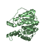
 Structure visualization
 Downloads & links
 Other downloads


 PDBj
PDBj


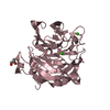
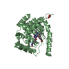
 Assembly
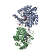


 Mass: 55.845 Da / Num. of mol.: 2 / Source method: obtained synthetically / Formula: Fe
 Mass: 122.143 Da / Num. of mol.: 2 / Source method: obtained synthetically / Formula: C4H12NO3 / Comment: pH buffer*YM
 Mass: 92.094 Da / Num. of mol.: 2 / Source method: obtained synthetically / Formula: C3H8O3
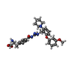
 Mass: 545.629 Da / Num. of mol.: 2 / Source method: obtained synthetically / Formula: C30H35N5O5
 Sample preparation
 / Beamline: 21-ID-F / Wavelength: 1 Å
 Processing