 Movie
Movie Controller

[English] 日本語
 Yorodumi
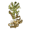
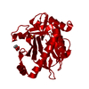
- PDB-5jqn: NitN Amidase from Neterenkonia sp. AN1 after thrombin His-tag removal. -
+ Open data

- Basic information
| Entry | Database: PDB / ID: 5jqn | ||||||
|---|---|---|---|---|---|---|---|
| Title | NitN Amidase from Neterenkonia sp. AN1 after thrombin His-tag removal. | ||||||
 Components Components | Aliphatic amidase | ||||||
 Keywords Keywords | HYDROLASE / NitN Amidase / Neterenkonia sp. AN1 | ||||||
| Function / homology |  Function and homology information Function and homology information | ||||||
| Biological species |  Nesterenkonia sp. AN1 (bacteria) Nesterenkonia sp. AN1 (bacteria) | ||||||
| Method |  X-RAY DIFFRACTION / SYNCHROTRON / MOLECULAR REPLACEMENT / Resolution: 1.19 Å X-RAY DIFFRACTION / SYNCHROTRON / MOLECULAR REPLACEMENT / Resolution: 1.19 Å | ||||||
 Authors Authors | Sewell, B.T. / Kimani, S.W. / Weber, B.W. | ||||||
 Citation Citation | Journal: To Be Published Title: QM/MM Modelling of Substrate Binding in the Amidase Active Site Authors: Sewell, B.T. / Kimani, S.W. / Venter, G.A. / Hunter, R. / Schell, D.T. #1: Journal: Appl. Environ. Microbiol. / Year: 2011Title: Unique aliphatic amidase from a psychrotrophic and haloalkaliphilic nesterenkonia isolate. Authors: Nel, A.J. / Tuffin, I.M. / Sewell, B.T. / Cowan, D.A. | ||||||
| History |
|
- Structure visualization
| Structure viewer | Molecule: MolmilJmol/JSmol |
|---|
- Downloads & links
-Download
| PDBx/mmCIF format | 5jqn.cif.gz | 68.2 KB | Display | PDBx/mmCIF format |
|---|---|---|---|---|
| PDB format | pdb5jqn.ent.gz | 48.6 KB | Display | PDB format |
| PDBx/mmJSON format | 5jqn.json.gz | Tree view | PDBx/mmJSON format | |
| Others |  Other downloads Other downloads |
-Validation report
| Arichive directory | https://data.pdbj.org/pub/pdb/validation_reports/jq/5jqnftp://data.pdbj.org/pub/pdb/validation_reports/jq/5jqn | HTTPS FTP |
|---|
-Related structure data
| Related structure data |  3hkxS S: Starting model for refinement |
|---|---|
| Similar structure data |


-Links
 PDBj
PDBj- Assembly
| Deposited unit | 
| |||||||||||||||
|---|---|---|---|---|---|---|---|---|---|---|---|---|---|---|---|---|
| 1 | 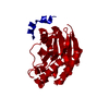
| |||||||||||||||
| Unit cell |
| |||||||||||||||
| Components on special symmetry positions |
|
-Components
| #1: Protein | Mass: 28243.664 Da / Num. of mol.: 1 / Mutation: GSH arising from the N-terminal His-tag. Source method: isolated from a genetically manipulated source Source: (gene. exp.) Nesterenkonia sp. AN1 (bacteria) / Gene: nit2 / Production host: |
|---|---|
| #2: Water | ChemComp-HOH /  Mass: 18.015 Da / Num. of mol.: 191 / Source method: isolated from a natural source / Formula: H2O Mass: 18.015 Da / Num. of mol.: 191 / Source method: isolated from a natural source / Formula: H2O |
| Has protein modification | Y |
-Experimental details
-Experiment
| Experiment | Method: X-RAY DIFFRACTION / Number of used crystals: 1 |
|---|
- Sample preparation
| Crystal | Density Matthews: 2.54 Å3/Da / Density % sol: 51.61 % |
|---|---|
| Crystal grow | Temperature: 295.15 K / Method: vapor diffusion, sitting drop / pH: 7.5 / Details: 2M Ammonium Sufate |
-Data collection
| Diffraction | Mean temperature: 100 K |
|---|---|
| Diffraction source | Source: SYNCHROTRON / Site: Diamond  / Beamline: I04-1 / Wavelength: 0.9173 Å / Beamline: I04-1 / Wavelength: 0.9173 Å |
| Detector | Type: DECTRIS PILATUS 2M / Detector: PIXEL / Date: Mar 8, 2015 / Details: Toroidal Mirror |
| Radiation | Protocol: SINGLE WAVELENGTH / Monochromatic (M) / Laue (L): M / Scattering type: x-ray |
| Radiation wavelength | Wavelength: 0.9173 Å / Relative weight: 1 |
| Reflection | Resolution: 1.19→45.566 Å / Num. obs: 91215 / % possible obs: 98.6 % / Redundancy: 5.6 % / Rrim(I) all: 0.037 / Net I/σ(I): 25.13 / Num. measured all: 580951 |
| Reflection shell | Highest resolution: 1.19 Å |
- Processing
| Software |
| |||||||||||||||||||||||||||||||||||||||||||||||||||||||||||||||||||||||||||||||||||||||||||||||||||||||||
|---|---|---|---|---|---|---|---|---|---|---|---|---|---|---|---|---|---|---|---|---|---|---|---|---|---|---|---|---|---|---|---|---|---|---|---|---|---|---|---|---|---|---|---|---|---|---|---|---|---|---|---|---|---|---|---|---|---|---|---|---|---|---|---|---|---|---|---|---|---|---|---|---|---|---|---|---|---|---|---|---|---|---|---|---|---|---|---|---|---|---|---|---|---|---|---|---|---|---|---|---|---|---|---|---|---|---|
| Refinement | Method to determine structure: MOLECULAR REPLACEMENT Starting model: 3HKX Resolution: 1.19→45.566 Å / SU ML: 0.1 / Cross valid method: FREE R-VALUE / σ(F): 1.34 / Phase error: 18.08 / Stereochemistry target values: ML
| |||||||||||||||||||||||||||||||||||||||||||||||||||||||||||||||||||||||||||||||||||||||||||||||||||||||||
| Solvent computation | Shrinkage radii: 0.9 Å / VDW probe radii: 1.11 Å / Solvent model: FLAT BULK SOLVENT MODEL | |||||||||||||||||||||||||||||||||||||||||||||||||||||||||||||||||||||||||||||||||||||||||||||||||||||||||
| Refinement step | Cycle: LAST / Resolution: 1.19→45.566 Å
| |||||||||||||||||||||||||||||||||||||||||||||||||||||||||||||||||||||||||||||||||||||||||||||||||||||||||
| Refine LS restraints |
| |||||||||||||||||||||||||||||||||||||||||||||||||||||||||||||||||||||||||||||||||||||||||||||||||||||||||
| LS refinement shell |
|
