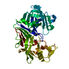
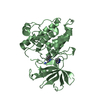
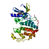
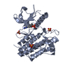
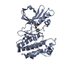
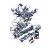
Entry Database : PDB / ID : 4xeyTitle Crystal structure of an SH2-kinase domain construct of c-Abl tyrosine kinase Tyrosine-protein kinase ABL1 Keywords / / / / / Function / homology Function Domain/homology Component
/ / / / / / / / / / / / / / / / / / / / / / / / / / / / / / / / / / / / / / / / / / / / / / / / / / / / / / / / / / / / / / / / / / / / / / / / / / / / / / / / / / / / / / / / / / / / / / / / / / / / / / / / / / / / / / / / / / / / / / / / / / / / / / / / / / / / / / / / / / / / / / / / / / / / / / Biological species Homo sapiens (human)Method / / / Resolution : 2.891 Å Authors Lorenz, S. / Deng, P. / Kuriyan, J. Funding support Organization Grant number Country Leukemia & Lymphoma Society 7393-06
Journal : Biochem.J. / Year : 2015Title : Crystal structure of an SH2-kinase construct of c-Abl and effect of the SH2 domain on kinase activity.Authors : Lorenz, S. / Deng, P. / Hantschel, O. / Superti-Furga, G. / Kuriyan, J. History Deposition Dec 25, 2014 Deposition site / Processing site Revision 1.0 Apr 1, 2015 Provider / Type Revision 1.1 Jun 3, 2015 Group Revision 1.2 Sep 27, 2017 Group Author supporting evidence / Database references ... Author supporting evidence / Database references / Derived calculations / Source and taxonomy Category citation / entity_src_gen ... citation / entity_src_gen / pdbx_audit_support / pdbx_struct_oper_list Item _citation.journal_id_CSD / _entity_src_gen.pdbx_alt_source_flag ... _citation.journal_id_CSD / _entity_src_gen.pdbx_alt_source_flag / _pdbx_audit_support.funding_organization / _pdbx_struct_oper_list.symmetry_operation Revision 1.3 Sep 27, 2023 Group / Database references / Refinement descriptionCategory chem_comp_atom / chem_comp_bond ... chem_comp_atom / chem_comp_bond / database_2 / pdbx_initial_refinement_model Item / _database_2.pdbx_database_accession
Show all Show less
 Movie
Movie Controller

 Yorodumi
 Open data
 Basic information
 Components
 Keywords
 Function and homology information
 Homo sapiens (human)
 X-RAY DIFFRACTION /
  Authors
 United States, 1items
  Citation
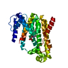
 Structure visualization
 Downloads & links
 Other downloads
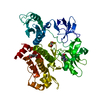


 PDBj
PDBj


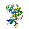
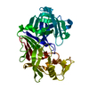
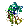
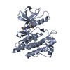
 Assembly
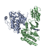


 Mass: 488.006 Da / Num. of mol.: 2 / Source method: obtained synthetically / Formula: C22H26ClN7O2S / Comment: medication*YM
 Sample preparation
 Processing