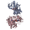
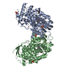
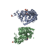
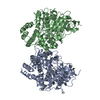
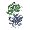
- PDB-5kvh: Crystal structure of human apoptosis-inducing factor with W196A m... -
+
Open data
ID or keywords:
Loading...
-
Basic information
Entry
Database: PDB / ID: 5kvh
Title
Crystal structure of human apoptosis-inducing factor with W196A mutation
Components
Apoptosis-inducing factor 1, mitochondrial
Keywords
OXIDOREDUCTASE / Flavoprotein / Mitochondria / Cell death
Function / homology
Function and homology information
Oxidoreductases; Acting on NADH or NADPH; With unknown physiological acceptors / protein import into the intermembrane space via the disulfide relay system / cellular response to aldosterone / mitochondrial respiratory chain complex assembly / protein import into mitochondrial intermembrane space / poly-ADP-D-ribose binding / NAD(P)H oxidase H2O2-forming activity / positive regulation of necroptotic process / response to L-glutamate / intrinsic apoptotic signaling pathway in response to endoplasmic reticulum stress ...Oxidoreductases; Acting on NADH or NADPH; With unknown physiological acceptors / protein import into the intermembrane space via the disulfide relay system / cellular response to aldosterone / mitochondrial respiratory chain complex assembly / protein import into mitochondrial intermembrane space / poly-ADP-D-ribose binding / NAD(P)H oxidase H2O2-forming activity / positive regulation of necroptotic process / response to L-glutamate / intrinsic apoptotic signaling pathway in response to endoplasmic reticulum stress / oxidoreductase activity, acting on NAD(P)H / NADH dehydrogenase activity / sperm head-tail coupling apparatus / cellular response to nitric oxide / FAD binding / response to ischemia / cellular response to estradiol stimulus / mitochondrial intermembrane space / response to toxic substance / cellular response to hydrogen peroxide / neuron differentiation / positive regulation of neuron apoptotic process / cellular response to hypoxia / protein dimerization activity / mitochondrial inner membrane / positive regulation of apoptotic process / apoptotic process / perinuclear region of cytoplasm / mitochondrion / DNA binding / nucleus / cytosol Similarity search - Function
Apoptosis-inducingfactor1, mitochondrial / Programmed cell death protein 8
Mass: 59355.434 Da / Num. of mol.: 2 / Mutation: W196A Source method: isolated from a genetically manipulated source Details: Expressed as AIF(78-613). Residues L614-Q619 remain from Prescission protease cleavage. Source: (gene. exp.) Homo sapiens (human) / Gene: AIFM1, AIF, PDCD8 / Plasmid: pET24b / Production host: Escherichia coli (E. coli) / Strain (production host): Rosetta2 (DE3) References: UniProt: O95831, Oxidoreductases; Acting on the CH-OH group of donors; With NAD+ or NADP+ as acceptor
Resolution: 2.273→60.822 Å / SU ML: 0.35 / Cross valid method: FREE R-VALUE / σ(F): 1.33 / Phase error: 26.42 / Stereochemistry target values: ML Details: Hydrogens were included in riding positions during refinement. FAD cofactor and glycerol geometry restraints were prepared in eLBOW using Mogul or simple optimization, respectively, and ...Details: Hydrogens were included in riding positions during refinement. FAD cofactor and glycerol geometry restraints were prepared in eLBOW using Mogul or simple optimization, respectively, and included explicitly with the refinement. Residues 78-125, 511-559, and 613-619 are disordered in chains A and B.
Rfactor
Num. reflection
% reflection
Rfree
0.2395
2824
4.95 %
Rwork
0.2022
-
-
obs
0.204
57018
99.15 %
Solvent computation
Shrinkage radii: 0.9 Å / VDW probe radii: 1.11 Å / Solvent model: FLAT BULK SOLVENT MODEL
Refinement step
Cycle: LAST / Resolution: 2.273→60.822 Å
Protein
Nucleic acid
Ligand
Solvent
Total
Num. atoms
6699
0
112
683
7494
Refine LS restraints
Refine-ID
Type
Dev ideal
Number
X-RAY DIFFRACTION
f_bond_d
0.002
7033
X-RAY DIFFRACTION
f_angle_d
0.437
9515
X-RAY DIFFRACTION
f_dihedral_angle_d
8.117
4170
X-RAY DIFFRACTION
f_chiral_restr
0.045
1048
X-RAY DIFFRACTION
f_plane_restr
0.002
1212
LS refinement shell
Resolution (Å)
Rfactor Rfree
Num. reflection Rfree
Rfactor Rwork
Num. reflection Rwork
Refine-ID
% reflection obs (%)
2.2732-2.3124
0.3307
109
0.3286
2400
X-RAY DIFFRACTION
88
2.3124-2.3544
0.3345
139
0.3095
2690
X-RAY DIFFRACTION
100
2.3544-2.3997
0.3654
130
0.3
2688
X-RAY DIFFRACTION
100
2.3997-2.4487
0.3166
141
0.2901
2699
X-RAY DIFFRACTION
100
2.4487-2.5019
0.3543
115
0.2876
2692
X-RAY DIFFRACTION
100
2.5019-2.5601
0.3045
131
0.2654
2739
X-RAY DIFFRACTION
100
2.5601-2.6241
0.3106
131
0.258
2693
X-RAY DIFFRACTION
100
2.6241-2.6951
0.2955
149
0.2928
2678
X-RAY DIFFRACTION
99
2.6951-2.7744
0.2704
149
0.2493
2715
X-RAY DIFFRACTION
100
2.7744-2.864
0.2998
157
0.2356
2710
X-RAY DIFFRACTION
100
2.864-2.9663
0.2886
137
0.2163
2695
X-RAY DIFFRACTION
100
2.9663-3.0851
0.2536
155
0.2119
2701
X-RAY DIFFRACTION
100
3.0851-3.2255
0.2364
146
0.1965
2728
X-RAY DIFFRACTION
100
3.2255-3.3955
0.2378
150
0.183
2721
X-RAY DIFFRACTION
100
3.3955-3.6082
0.2145
124
0.1847
2752
X-RAY DIFFRACTION
100
3.6082-3.8868
0.1922
155
0.171
2676
X-RAY DIFFRACTION
98
3.8868-4.2778
0.1784
162
0.1507
2709
X-RAY DIFFRACTION
99
4.2778-4.8966
0.176
159
0.1302
2758
X-RAY DIFFRACTION
100
4.8966-6.1683
0.2264
123
0.1792
2818
X-RAY DIFFRACTION
100
6.1683-60.8437
0.2532
162
0.2128
2932
X-RAY DIFFRACTION
100
Refinement TLS params.
Method: refined / Refine-ID: X-RAY DIFFRACTION
ID
L11 (°2)
L12 (°2)
L13 (°2)
L22 (°2)
L23 (°2)
L33 (°2)
S11 (Å °)
S12 (Å °)
S13 (Å °)
S21 (Å °)
S22 (Å °)
S23 (Å °)
S31 (Å °)
S32 (Å °)
S33 (Å °)
T11 (Å2)
T12 (Å2)
T13 (Å2)
T22 (Å2)
T23 (Å2)
T33 (Å2)
Origin x (Å)
Origin y (Å)
Origin z (Å)
1
4.386
-0.5943
-0.4138
2.4852
0.525
1.5304
0.1438
-0.0212
0.7266
-0.0075
-0.0124
-0.1012
-0.254
0.0312
-0.1462
0.3128
-0.0366
-0.0177
0.2863
-0.0583
0.4545
-5.699
22.1746
-36.0383
2
3.8692
-1.758
-0.4017
2.1736
0.337
0.4947
-0.051
-0.2219
0.0203
0.1357
0.0334
-0.3128
0.0699
0.0864
0.0149
0.3241
-0.039
-0.0209
0.3317
-0.0059
0.3926
11.1914
1.7672
-37.9751
3
2.553
-0.6135
-1.7444
1.9554
0.4893
4.1831
0.0025
0.1711
-0.0498
-0.1907
0.0041
0.0209
0.1077
-0.0469
0.0191
0.2978
-0.0497
-0.0349
0.3175
-0.0178
0.3046
-8.9306
3.5548
-51.7212
Refinement TLS group
ID
Refine-ID
Refine TLS-ID
Selection details
1
X-RAY DIFFRACTION
1
chain 'A' and (resid129through245 )
2
X-RAY DIFFRACTION
2
chain 'A' and (resid246through402 )
3
X-RAY DIFFRACTION
3
chain 'A' and (resid403through611 )
+
About Yorodumi
-
News
-
Feb 9, 2022. New format data for meta-information of EMDB entries
New format data for meta-information of EMDB entries
Version 3 of the EMDB header file is now the official format.
The previous official version 1.9 will be removed from the archive.
In the structure databanks used in Yorodumi, some data are registered as the other names, "COVID-19 virus" and "2019-nCoV". Here are the details of the virus and the list of structure data.
Jan 31, 2019. EMDB accession codes are about to change! (news from PDBe EMDB page)
EMDB accession codes are about to change! (news from PDBe EMDB page)
The allocation of 4 digits for EMDB accession codes will soon come to an end. Whilst these codes will remain in use, new EMDB accession codes will include an additional digit and will expand incrementally as the available range of codes is exhausted. The current 4-digit format prefixed with “EMD-” (i.e. EMD-XXXX) will advance to a 5-digit format (i.e. EMD-XXXXX), and so on. It is currently estimated that the 4-digit codes will be depleted around Spring 2019, at which point the 5-digit format will come into force.
The EM Navigator/Yorodumi systems omit the EMD- prefix.
Related info.:Q: What is EMD? / ID/Accession-code notation in Yorodumi/EM Navigator
Yorodumi is a browser for structure data from EMDB, PDB, SASBDB, etc.
This page is also the successor to EM Navigator detail page, and also detail information page/front-end page for Omokage search.
The word "yorodu" (or yorozu) is an old Japanese word meaning "ten thousand". "mi" (miru) is to see.
Related info.:EMDB / PDB / SASBDB / Comparison of 3 databanks / Yorodumi Search / Aug 31, 2016. New EM Navigator & Yorodumi / Yorodumi Papers / Jmol/JSmol / Function and homology information / Changes in new EM Navigator and Yorodumi
 Movie
Movie Controller

 Yorodumi
 Open data
 Basic information
 Components
 Keywords
 Function and homology information
 Homo sapiens (human)
 X-RAY DIFFRACTION /
  Authors
 United States, 2items
  Citation
 Structure visualization
 Downloads & links
 Other downloads


 PDBj
PDBj


 Assembly
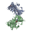


 Mass: 785.550 Da / Num. of mol.: 2 / Source method: obtained synthetically / Formula: C27H33N9O15P2 / Comment: FAD*YM
 Mass: 92.094 Da / Num. of mol.: 1 / Source method: obtained synthetically / Formula: C3H8O3
 Mass: 18.015 Da / Num. of mol.: 683 / Source method: isolated from a natural source / Formula: H2O
 Sample preparation
 Processing