- PDB-5miv: G307E variant of murine Apoptosis Inducing Factor in complex with NAD+ -
+
Open data
ID or keywords:
Loading...
-
Basic information
Entry
Database: PDB / ID: 5miv
Title
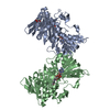
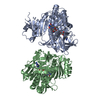
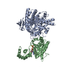
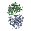
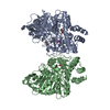
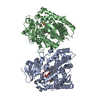
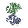
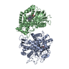
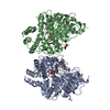
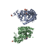
G307E variant of murine Apoptosis Inducing Factor in complex with NAD+
Components
Apoptosis-inducing factor 1, mitochondrial
Keywords
OXIDOREDUCTASE / Apoptosis Inducing Factor
Function / homology
Function and homology information
regulation of apoptotic DNA fragmentation / electron-transferring-flavoprotein dehydrogenase activity / Oxidoreductases; Acting on NADH or NADPH; With unknown physiological acceptors / protein import into the intermembrane space via the disulfide relay system / mitochondrial respiratory chain complex assembly / protein import into mitochondrial intermembrane space / poly-ADP-D-ribose binding / NAD(P)H oxidase H2O2-forming activity / positive regulation of necroptotic process / apoptotic mitochondrial changes ...regulation of apoptotic DNA fragmentation / electron-transferring-flavoprotein dehydrogenase activity / Oxidoreductases; Acting on NADH or NADPH; With unknown physiological acceptors / protein import into the intermembrane space via the disulfide relay system / mitochondrial respiratory chain complex assembly / protein import into mitochondrial intermembrane space / poly-ADP-D-ribose binding / NAD(P)H oxidase H2O2-forming activity / positive regulation of necroptotic process / apoptotic mitochondrial changes / intrinsic apoptotic signaling pathway in response to endoplasmic reticulum stress / oxidoreductase activity, acting on NAD(P)H / NADH dehydrogenase activity / FAD binding / mitochondrial intermembrane space / neuron differentiation / positive regulation of neuron apoptotic process / neuron apoptotic process / response to oxidative stress / mitochondrial outer membrane / protein dimerization activity / mitochondrial inner membrane / positive regulation of apoptotic process / apoptotic process / perinuclear region of cytoplasm / mitochondrion / DNA binding / nucleus / cytosol / cytoplasm Similarity search - Function
Component-ID: _ / Ens-ID: 1 / Beg auth comp-ID: PRO / Beg label comp-ID: PRO / End auth comp-ID: GLU / End label comp-ID: GLU / Refine code: _ / Auth seq-ID: 128 - 611 / Label seq-ID: 51 - 534
Dom-ID
Auth asym-ID
Label asym-ID
1
A
A
2
C
B
-
Components
#1: Protein
Apoptosis-inducingfactor1, mitochondrial / Programmed cell death protein 8
Mass: 58400.266 Da / Num. of mol.: 2 Source method: isolated from a genetically manipulated source Source: (gene. exp.) Mus musculus (house mouse) / Gene: Aifm1, Aif, Pdcd8 / Production host: Escherichia coli BL21(DE3) (bacteria) References: UniProt: Q9Z0X1, Oxidoreductases; Acting on the CH-OH group of donors; With NAD+ or NADP+ as acceptor
Resolution: 3.1→43.5 Å / Cor.coef. Fo:Fc: 0.956 / Cor.coef. Fo:Fc free: 0.914 / Cross valid method: THROUGHOUT / ESU R Free: 0.442 / Details: HYDROGENS HAVE BEEN ADDED IN THE RIDING POSITIONS
Rfactor
Num. reflection
% reflection
Selection details
Rfree
0.26269
1322
5 %
RANDOM
Rwork
0.19795
-
-
-
obs
0.20121
25254
99.52 %
-
Solvent computation
Ion probe radii: 0.8 Å / Shrinkage radii: 0.8 Å / VDW probe radii: 1.2 Å
 Movie
Movie Controller

 Yorodumi
 Open data
 Basic information
 Components
 Keywords
 Function and homology information
 X-RAY DIFFRACTION /
  Authors
 Citation
 Structure visualization
 Downloads & links
 Other downloads

 PDBj
PDBj


 Assembly
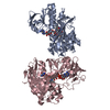


 Mass: 663.425 Da / Num. of mol.: 2 / Source method: obtained synthetically / Formula: C21H27N7O14P2 / Comment: NAD*YM
 Mass: 785.550 Da / Num. of mol.: 2 / Source method: obtained synthetically / Formula: C27H33N9O15P2 / Comment: FAD*YM
 Mass: 18.015 Da / Num. of mol.: 125 / Source method: isolated from a natural source / Formula: H2O
 Sample preparation
 / Beamline: ID29 / Wavelength: 0.97895 Å
 Processing