 Movie
Movie Controller

[English] 日本語
 Yorodumi
- PDB-3b98: Crystal structure of zebrafish prostacyclin synthase (cytochrome ... -
+ Open data

- Basic information
| Entry | Database: PDB / ID: 3b98 | ||||||
|---|---|---|---|---|---|---|---|
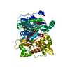
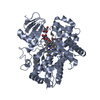
| Title | Crystal structure of zebrafish prostacyclin synthase (cytochrome P450 8A1) | ||||||
 Components Components | Prostaglandin I2 synthase | ||||||
 Keywords Keywords | ISOMERASE / prostacyclin synthase / cytochrome p450 8A1 / CYP8A1 | ||||||
| Function / homology |  Function and homology information Function and homology informationEicosanoids / Synthesis of Prostaglandins (PG) and Thromboxanes (TX) / prostaglandin-I synthase / prostaglandin-I synthase activity / hydroperoxy icosatetraenoate dehydratase / hydroperoxy icosatetraenoate dehydratase activity / prostaglandin biosynthetic process / oxidoreductase activity, acting on paired donors, with incorporation or reduction of molecular oxygen / monooxygenase activity / iron ion binding ...Eicosanoids / Synthesis of Prostaglandins (PG) and Thromboxanes (TX) / prostaglandin-I synthase / prostaglandin-I synthase activity / hydroperoxy icosatetraenoate dehydratase / hydroperoxy icosatetraenoate dehydratase activity / prostaglandin biosynthetic process / oxidoreductase activity, acting on paired donors, with incorporation or reduction of molecular oxygen / monooxygenase activity / iron ion binding / heme binding / endoplasmic reticulum membrane Similarity search - Function | ||||||
| Biological species |  | ||||||
| Method |  X-RAY DIFFRACTION / SYNCHROTRON / MOLECULAR REPLACEMENT / Resolution: 2.08 Å X-RAY DIFFRACTION / SYNCHROTRON / MOLECULAR REPLACEMENT / Resolution: 2.08 Å | ||||||
 Authors Authors | Li, Y.-C. / Chiang, C.-W. / Yeh, H.-C. / Hsu, P.-Y. / Whitby, F.G. / Wang, L.-H. / Chan, N.-L. | ||||||
 Citation Citation | Journal: J.Biol.Chem. / Year: 2008 Title: Structures of Prostacyclin Synthase and Its Complexes with Substrate Analog and Inhibitor Reveal a Ligand-specific Heme Conformation Change Authors: Li, Y.-C. / Chiang, C.-W. / Yeh, H.-C. / Hsu, P.-Y. / Whitby, F.G. / Wang, L.-H. / Chan, N.-L. | ||||||
| History |
|
- Structure visualization
| Structure viewer | Molecule: MolmilJmol/JSmol |
|---|
- Downloads & links
-Download
| PDBx/mmCIF format | 3b98.cif.gz | 195 KB | Display | PDBx/mmCIF format |
|---|---|---|---|---|
| PDB format | pdb3b98.ent.gz | 154 KB | Display | PDB format |
| PDBx/mmJSON format | 3b98.json.gz | Tree view | PDBx/mmJSON format | |
| Others |  Other downloads Other downloads |
-Validation report
| Arichive directory | https://data.pdbj.org/pub/pdb/validation_reports/b9/3b98ftp://data.pdbj.org/pub/pdb/validation_reports/b9/3b98 | HTTPS FTP |
|---|
-Related structure data
| Related structure data |  3b6hC  3b99C  2iagS C: citing same article ( S: Starting model for refinement |
|---|---|
| Similar structure data |


-Links
 PDBj
PDBj
- Assembly
| Deposited unit | 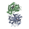
| ||||||||
|---|---|---|---|---|---|---|---|---|---|
| 1 | 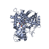
| ||||||||
| 2 | 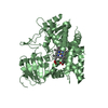
| ||||||||
| Unit cell |
|
-Components
| #1: Protein | Mass: 54848.930 Da / Num. of mol.: 2 / Fragment: UNP residues 17-480 Source method: isolated from a genetically manipulated source Source: (gene. exp.)  References: UniProt: A9LLA5, UniProt: F1RE08*PLUS, prostaglandin-I synthase #2: Chemical |   Mass: 616.487 Da / Num. of mol.: 2 / Source method: obtained synthetically / Formula: C34H32FeN4O4 Mass: 616.487 Da / Num. of mol.: 2 / Source method: obtained synthetically / Formula: C34H32FeN4O4#3: Water | ChemComp-HOH / |  Mass: 18.015 Da / Num. of mol.: 282 / Source method: isolated from a natural source / Formula: H2O Mass: 18.015 Da / Num. of mol.: 282 / Source method: isolated from a natural source / Formula: H2O |
|---|
-Experimental details
-Experiment
| Experiment | Method: X-RAY DIFFRACTION / Number of used crystals: 1 |
|---|
- Sample preparation
| Crystal | Density Matthews: 2.24 Å3/Da / Density % sol: 45.16 % |
|---|---|
| Crystal grow | Temperature: 277 K / Method: vapor diffusion, hanging drop / pH: 8 Details: 1 microliter of concentrated protein solution (20mg/ml) in gel filtration buffer (20mM Tris-HCl pH 8.0, 150mM NaCl and 5mM beta-mercaptoethanol) was mixed with equal amount of reservoir ...Details: 1 microliter of concentrated protein solution (20mg/ml) in gel filtration buffer (20mM Tris-HCl pH 8.0, 150mM NaCl and 5mM beta-mercaptoethanol) was mixed with equal amount of reservoir solution (20% PEG 3350 plus either 50mM Tris-HCl (pH 8.0) or 50mM HEPES (Na-salt; pH 7.5)) and equilibrated against 450 microliter of reservoir solution at 277 K, VAPOR DIFFUSION, HANGING DROP |
-Data collection
| Diffraction | Mean temperature: 100 K |
|---|---|
| Diffraction source | Source: SYNCHROTRON / Site: NSRRC  / Beamline: BL13B1 / Wavelength: 1 Å / Beamline: BL13B1 / Wavelength: 1 Å |
| Detector | Type: ADSC QUANTUM 315 / Detector: CCD / Date: Dec 13, 2006 / Details: Toroidal FM |
| Radiation | Monochromator: Si(111) / Protocol: SINGLE WAVELENGTH / Monochromatic (M) / Laue (L): M / Scattering type: x-ray |
| Radiation wavelength | Wavelength: 1 Å / Relative weight: 1 |
| Reflection | Resolution: 2.08→30 Å / Num. all: 60147 / Num. obs: 60087 / % possible obs: 99.9 % / Observed criterion σ(F): 1 / Observed criterion σ(I): 1 / Redundancy: 5.4 % / Rsym value: 0.046 / Net I/σ(I): 18 |
| Reflection shell | Resolution: 2.08→2.15 Å / Redundancy: 5.4 % / Mean I/σ(I) obs: 4.3 / Num. unique all: 5912 / Rsym value: 0.21 / % possible all: 99.8 |
- Processing
| Software |
| ||||||||||||||||||||||||||||||||||||||||||||||||||||||||||||||||||||||||||||||||||||||||||
|---|---|---|---|---|---|---|---|---|---|---|---|---|---|---|---|---|---|---|---|---|---|---|---|---|---|---|---|---|---|---|---|---|---|---|---|---|---|---|---|---|---|---|---|---|---|---|---|---|---|---|---|---|---|---|---|---|---|---|---|---|---|---|---|---|---|---|---|---|---|---|---|---|---|---|---|---|---|---|---|---|---|---|---|---|---|---|---|---|---|---|---|
| Refinement | Method to determine structure: MOLECULAR REPLACEMENT Starting model: PDB ENTRY 2IAG Resolution: 2.08→30 Å / Cor.coef. Fo:Fc: 0.924 / Cor.coef. Fo:Fc free: 0.895 / SU B: 4.904 / SU ML: 0.137 / Cross valid method: THROUGHOUT / σ(F): 1 / σ(I): 1 / ESU R: 0.254 / ESU R Free: 0.203 / Stereochemistry target values: MAXIMUM LIKELIHOOD / Details: HYDROGENS HAVE BEEN ADDED IN THE RIDING POSITIONS
| ||||||||||||||||||||||||||||||||||||||||||||||||||||||||||||||||||||||||||||||||||||||||||
| Solvent computation | Ion probe radii: 0.8 Å / Shrinkage radii: 0.8 Å / VDW probe radii: 1.4 Å / Solvent model: MASK | ||||||||||||||||||||||||||||||||||||||||||||||||||||||||||||||||||||||||||||||||||||||||||
| Displacement parameters | Biso mean: 28.442 Å2
| ||||||||||||||||||||||||||||||||||||||||||||||||||||||||||||||||||||||||||||||||||||||||||
| Refinement step | Cycle: LAST / Resolution: 2.08→30 Å
| ||||||||||||||||||||||||||||||||||||||||||||||||||||||||||||||||||||||||||||||||||||||||||
| Refine LS restraints |
| ||||||||||||||||||||||||||||||||||||||||||||||||||||||||||||||||||||||||||||||||||||||||||
| LS refinement shell | Resolution: 2.081→2.135 Å / Total num. of bins used: 20
|
