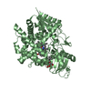
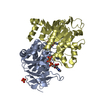
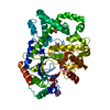
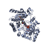
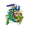
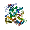
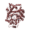
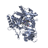
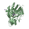
There are two prostacyclin synthase molecules in the asymmetric unit, but the enzyme should function as a monomer.
-
Components
#1: Protein
Prostacyclinsynthase / Prostaglandin I2 synthase
Mass: 55355.395 Da / Num. of mol.: 2 / Fragment: residues 23-500 Source method: isolated from a genetically manipulated source Source: (gene. exp.) Homo sapiens (human) Description: Plasmid pCW was a gift from Dr. Amy Roth, University of Oregon. Gene: PTGIS / Plasmid: pCW / Production host: Escherichia coli (E. coli) / Strain (production host): BL21(DE3), B834(DE3) / References: UniProt: Q16647, prostaglandin-I synthase
Mass: 18.015 Da / Num. of mol.: 435 / Source method: isolated from a natural source / Formula: H2O
-
Experimental details
-
Experiment
Experiment
Method: X-RAY DIFFRACTION / Number of used crystals: 2
-
Sample preparation
Crystal
Density Matthews: 2.39 Å3/Da / Density % sol: 48.45 %
Crystal grow
Temperature: 277 K / Method: vapor diffusion, hanging drop / pH: 8 Details: The native recombinant PGIS (25 mg/ml in the buffer containing 20 mM Tris, pH 8.0 and 150 mM NaCl) was found to crystallize spontaneously into needle clusters in the Eppendorf tube when ...Details: The native recombinant PGIS (25 mg/ml in the buffer containing 20 mM Tris, pH 8.0 and 150 mM NaCl) was found to crystallize spontaneously into needle clusters in the Eppendorf tube when stored at 4 C for about a week. A combination of microseeding technique and the hanging-drop vapor-diffusion method was then used to improve crystal quality. Specifically, seed stock was prepared by crushing these initial PGIS needle clusters with the Seed Bead Kit (Hampton Research). Following four 10-fold serial dilutions of the seed stock using the gel filtration buffer, 1 micro-l of diluted seeds was added into a 10 micro-l drop of freshly concentrated PGIS sample, and equilibrated at 4 C against 450 micro-l of the gel filtration buffer. High quality single crystals of PGIS were most often obtained using 100- or 1000-fold diluted seed stock, and reached suitable size (~ 0.05,0.05 ~ 0.1 mm3) within one week., VAPOR DIFFUSION, HANGING DROP, temperature 277K
-
Data collection
Diffraction
ID
Mean temperature (K)
Crystal-ID
1
100
1
2
100
1
Diffraction source
Source
Site
Beamline
ID
Wavelength (Å)
SYNCHROTRON
NSRRC
BL13B1
1
0.980127
SYNCHROTRON
SPring-8
BL12B2
2
0.9537, 0.9793
Detector
Type
ID
Detector
Date
ADSC QUANTUM 315
1
CCD
Jun 29, 2006
ADSC QUANTUM 4
2
CCD
May 15, 2006
Radiation
ID
Protocol
Monochromatic (M) / Laue (L)
Scattering type
Wavelength-ID
1
SINGLEWAVELENGTH
M
x-ray
1
2
MAD
M
x-ray
1
Radiation wavelength
ID
Wavelength (Å)
Relative weight
1
0.980127
1
2
0.9537
1
3
0.9793
1
Reflection
Resolution: 2.15→30 Å / Num. all: 56459 / Num. obs: 51322 / % possible obs: 90.9 % / Redundancy: 2.8 % / Rsym value: 0.063 / Net I/σ(I): 12.7
Reflection shell
Resolution: 2.15→2.23 Å / Mean I/σ(I) obs: 3.1 / Rsym value: 0.35 / % possible all: 93.5
-
Processing
Software
Name
Version
Classification
REFMAC
5.2.0019
refinement
HKL-2000
datacollection
HKL-2000
datareduction
HKL-2000
datascaling
SOLVE
phasing
Refinement
Method to determine structure: MAD / Resolution: 2.15→20.15 Å / Cor.coef. Fo:Fc: 0.938 / Cor.coef. Fo:Fc free: 0.906 / SU B: 5.798 / SU ML: 0.155 / Cross valid method: THROUGHOUT / ESU R: 0.322 / ESU R Free: 0.243 / Stereochemistry target values: MAXIMUM LIKELIHOOD / Details: HYDROGENS HAVE BEEN ADDED IN THE RIDING POSITIONS
Rfactor
Num. reflection
% reflection
Selection details
Rfree
0.26313
2601
5.1 %
RANDOM
Rwork
0.2009
-
-
-
obs
0.20407
48702
90.96 %
-
Solvent computation
Ion probe radii: 0.8 Å / Shrinkage radii: 0.8 Å / VDW probe radii: 1.4 Å / Solvent model: MASK
Displacement parameters
Biso mean: 26.948 Å2
Baniso -1
Baniso -2
Baniso -3
1-
0 Å2
0 Å2
0 Å2
2-
-
0 Å2
0 Å2
3-
-
-
0 Å2
Refinement step
Cycle: LAST / Resolution: 2.15→20.15 Å
Protein
Nucleic acid
Ligand
Solvent
Total
Num. atoms
7611
0
87
435
8133
Refine LS restraints
Refine-ID
Type
Dev ideal
Dev ideal target
Number
X-RAY DIFFRACTION
r_bond_refined_d
0.011
0.022
7915
X-RAY DIFFRACTION
r_bond_other_d
X-RAY DIFFRACTION
r_angle_refined_deg
1.38
2.001
10757
X-RAY DIFFRACTION
r_angle_other_deg
X-RAY DIFFRACTION
r_dihedral_angle_1_deg
6.177
5
935
X-RAY DIFFRACTION
r_dihedral_angle_2_deg
32.256
22.433
374
X-RAY DIFFRACTION
r_dihedral_angle_3_deg
17.588
15
1325
X-RAY DIFFRACTION
r_dihedral_angle_4_deg
18.433
15
74
X-RAY DIFFRACTION
r_chiral_restr
0.095
0.2
1144
X-RAY DIFFRACTION
r_gen_planes_refined
0.005
0.02
6066
X-RAY DIFFRACTION
r_gen_planes_other
X-RAY DIFFRACTION
r_nbd_refined
0.205
0.2
3657
X-RAY DIFFRACTION
r_nbd_other
X-RAY DIFFRACTION
r_nbtor_refined
0.304
0.2
5280
X-RAY DIFFRACTION
r_nbtor_other
X-RAY DIFFRACTION
r_xyhbond_nbd_refined
0.16
0.2
480
X-RAY DIFFRACTION
r_xyhbond_nbd_other
X-RAY DIFFRACTION
r_metal_ion_refined
X-RAY DIFFRACTION
r_metal_ion_other
X-RAY DIFFRACTION
r_symmetry_vdw_refined
0.192
0.2
84
X-RAY DIFFRACTION
r_symmetry_vdw_other
X-RAY DIFFRACTION
r_symmetry_hbond_refined
0.154
0.2
25
X-RAY DIFFRACTION
r_symmetry_hbond_other
X-RAY DIFFRACTION
r_symmetry_metal_ion_refined
X-RAY DIFFRACTION
r_symmetry_metal_ion_other
X-RAY DIFFRACTION
r_mcbond_it
0.642
1.5
4689
X-RAY DIFFRACTION
r_mcbond_other
X-RAY DIFFRACTION
r_mcangle_it
1.213
2
7541
X-RAY DIFFRACTION
r_scbond_it
1.961
3
3373
X-RAY DIFFRACTION
r_scangle_it
2.987
4.5
3216
X-RAY DIFFRACTION
r_rigid_bond_restr
X-RAY DIFFRACTION
r_sphericity_free
X-RAY DIFFRACTION
r_sphericity_bonded
LS refinement shell
Resolution: 2.151→2.206 Å / Total num. of bins used: 20
Rfactor
Num. reflection
% reflection
Rfree
0.318
195
-
Rwork
0.215
3616
-
obs
-
-
93.29 %
+
About Yorodumi
-
News
-
Feb 9, 2022. New format data for meta-information of EMDB entries
New format data for meta-information of EMDB entries
Version 3 of the EMDB header file is now the official format.
The previous official version 1.9 will be removed from the archive.
In the structure databanks used in Yorodumi, some data are registered as the other names, "COVID-19 virus" and "2019-nCoV". Here are the details of the virus and the list of structure data.
Jan 31, 2019. EMDB accession codes are about to change! (news from PDBe EMDB page)
EMDB accession codes are about to change! (news from PDBe EMDB page)
The allocation of 4 digits for EMDB accession codes will soon come to an end. Whilst these codes will remain in use, new EMDB accession codes will include an additional digit and will expand incrementally as the available range of codes is exhausted. The current 4-digit format prefixed with “EMD-” (i.e. EMD-XXXX) will advance to a 5-digit format (i.e. EMD-XXXXX), and so on. It is currently estimated that the 4-digit codes will be depleted around Spring 2019, at which point the 5-digit format will come into force.
The EM Navigator/Yorodumi systems omit the EMD- prefix.
Related info.:Q: What is EMD? / ID/Accession-code notation in Yorodumi/EM Navigator
Yorodumi is a browser for structure data from EMDB, PDB, SASBDB, etc.
This page is also the successor to EM Navigator detail page, and also detail information page/front-end page for Omokage search.
The word "yorodu" (or yorozu) is an old Japanese word meaning "ten thousand". "mi" (miru) is to see.
Related info.:EMDB / PDB / SASBDB / Comparison of 3 databanks / Yorodumi Search / Aug 31, 2016. New EM Navigator & Yorodumi / Yorodumi Papers / Jmol/JSmol / Function and homology information / Changes in new EM Navigator and Yorodumi
 Movie
Movie Controller


 Open data
 Basic information
 Components
 Keywords
 Function and homology information
 Homo sapiens (human)
 X-RAY DIFFRACTION /
  Authors
 Citation
 Structure visualization
 Downloads & links
 Other downloads


 PDBj
PDBj

 Assembly
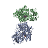


 Mass: 616.487 Da / Num. of mol.: 2 / Source method: obtained synthetically / Formula: C34H32FeN4O4
 Mass: 22.990 Da / Num. of mol.: 1 / Source method: obtained synthetically / Formula: Na
 Mass: 18.015 Da / Num. of mol.: 435 / Source method: isolated from a natural source / Formula: H2O
 Sample preparation

 Processing