 Movie
Movie Controller

[English] 日本語
 Yorodumi
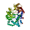
- PDB-6m6t: Amylomaltase from Streptococcus agalactiae in complex with acarbose -
+ Open data

- Basic information
| Entry | Database: PDB / ID: 6m6t | ||||||
|---|---|---|---|---|---|---|---|
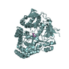
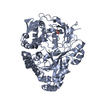
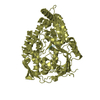
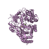
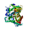
| Title | Amylomaltase from Streptococcus agalactiae in complex with acarbose | ||||||
 Components Components | 4-alpha-glucanotransferase | ||||||
 Keywords Keywords | TRANSFERASE / amylomaltase / 4-alpha-glucanotransferase / cyclodextrin / acarbose | ||||||
| Function / homology |  Function and homology information Function and homology information: / 4-alpha-glucanotransferase / 4-alpha-glucanotransferase activity / carbohydrate metabolic process Similarity search - Function | ||||||
| Biological species |  Streptococcus agalactiae (bacteria) Streptococcus agalactiae (bacteria) | ||||||
| Method |  X-RAY DIFFRACTION / SYNCHROTRON / MOLECULAR REPLACEMENT / molecular replacement / Resolution: 2.75 Å X-RAY DIFFRACTION / SYNCHROTRON / MOLECULAR REPLACEMENT / molecular replacement / Resolution: 2.75 Å | ||||||
 Authors Authors | Wangkanont, K. / Tumhom, S. / Pongsawasdi, P. | ||||||
 Citation Citation | Journal: Sci Rep / Year: 2021 Title: Streptococcus agalactiae amylomaltase offers insight into the transglycosylation mechanism and the molecular basis of thermostability among amylomaltases. Authors: Tumhom, S. / Nimpiboon, P. / Wangkanont, K. / Pongsawasdi, P. | ||||||
| History |
|
- Structure visualization
| Structure viewer | Molecule: MolmilJmol/JSmol |
|---|
- Downloads & links
-Download
| PDBx/mmCIF format | 6m6t.cif.gz | 796.4 KB | Display | PDBx/mmCIF format |
|---|---|---|---|---|
| PDB format | pdb6m6t.ent.gz | 667.6 KB | Display | PDB format |
| PDBx/mmJSON format | 6m6t.json.gz | Tree view | PDBx/mmJSON format | |
| Others |  Other downloads Other downloads |
-Validation report
| Arichive directory | https://data.pdbj.org/pub/pdb/validation_reports/m6/6m6tftp://data.pdbj.org/pub/pdb/validation_reports/m6/6m6t | HTTPS FTP |
|---|
-Related structure data
| Related structure data | 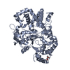 1tz7S S: Starting model for refinement |
|---|---|
| Similar structure data |

-Links
 PDBj
PDBj
- Assembly
| Deposited unit | 
| ||||||||
|---|---|---|---|---|---|---|---|---|---|
| 1 | 
| ||||||||
| 2 | 
| ||||||||
| 3 | 
| ||||||||
| 4 | 
| ||||||||
| 5 | 
| ||||||||
| 6 | 
| ||||||||
| 7 | 
| ||||||||
| 8 | 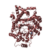
| ||||||||
| Unit cell |
|
-Components
| #1: Protein | Mass: 58983.957 Da / Num. of mol.: 8 / Fragment: amylomaltase, UNP residues 2-498 Source method: isolated from a genetically manipulated source Source: (gene. exp.) Streptococcus agalactiae (bacteria) / Gene: malQ / Plasmid: pET28a / Production host: #2: Polysaccharide | 4,6-dideoxy-4-{[(1S,4R,5S,6S)-4,5,6-trihydroxy-3-(hydroxymethyl)cyclohex-2-en-1-yl]amino}-alpha-D- ...4,6-dideoxy-4-{[(1S,4R,5S,6S)-4,5,6-trihydroxy-3-(hydroxymethyl)cyclohex-2-en-1-yl]amino}-alpha-D-glucopyranose-(1-4)-1,5-anhydro-D-glucitol Type: oligosaccharide / Mass: 467.465 Da / Num. of mol.: 7 Source method: isolated from a genetically manipulated source #3: Polysaccharide | 4,6-dideoxy-4-{[(1S,4R,5S,6S)-4,5,6-trihydroxy-3-(hydroxymethyl)cyclohex-2-en-1-yl]amino}-alpha-D- ...4,6-dideoxy-4-{[(1S,4R,5S,6S)-4,5,6-trihydroxy-3-(hydroxymethyl)cyclohex-2-en-1-yl]amino}-alpha-D-glucopyranose-(1-4)-alpha-D-glucopyranose / acarbose-derived trisaccharide 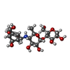  Type: oligosaccharide, Oligosaccharide / Class: Inhibitor / Mass: 483.465 Da / Num. of mol.: 6 Type: oligosaccharide, Oligosaccharide / Class: Inhibitor / Mass: 483.465 Da / Num. of mol.: 6Source method: isolated from a genetically manipulated source Details: oligosaccharide / References: acarbose-derived trisaccharide #4: Polysaccharide | 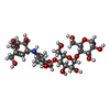  Type: oligosaccharide, Oligosaccharide / Class: Inhibitor / Mass: 645.606 Da / Num. of mol.: 3 Type: oligosaccharide, Oligosaccharide / Class: Inhibitor / Mass: 645.606 Da / Num. of mol.: 3Source method: isolated from a genetically manipulated source Details: oligosaccharide / References: alpha-acarbose #5: Water | ChemComp-HOH / |  Mass: 18.015 Da / Num. of mol.: 252 / Source method: isolated from a natural source / Formula: H2O Mass: 18.015 Da / Num. of mol.: 252 / Source method: isolated from a natural source / Formula: H2OHas ligand of interest | Y | |
|---|
-Experimental details
-Experiment
| Experiment | Method: X-RAY DIFFRACTION / Number of used crystals: 1 |
|---|
- Sample preparation
| Crystal | Density Matthews: 2.67 Å3/Da / Density % sol: 53.87 % / Mosaicity: 0.19 ° |
|---|---|
| Crystal grow | Temperature: 289 K / Method: batch mode / pH: 7.5 Details: 100 mM Tris, 100 mM Magnesium Formate, 15% PEG 8000 |
-Data collection
| Diffraction | Mean temperature: 100 K / Serial crystal experiment: N | ||||||||||||||||||||||||||||||
|---|---|---|---|---|---|---|---|---|---|---|---|---|---|---|---|---|---|---|---|---|---|---|---|---|---|---|---|---|---|---|---|
| Diffraction source | Source: SYNCHROTRON / Site: NSRRC  / Beamline: BL13B1 / Wavelength: 0.999999 Å / Beamline: BL13B1 / Wavelength: 0.999999 Å | ||||||||||||||||||||||||||||||
| Detector | Type: ADSC QUANTUM 315r / Detector: CCD / Date: Dec 21, 2019 | ||||||||||||||||||||||||||||||
| Radiation | Monochromator: Si(111) / Protocol: SINGLE WAVELENGTH / Monochromatic (M) / Laue (L): M / Scattering type: x-ray | ||||||||||||||||||||||||||||||
| Radiation wavelength | Wavelength: 0.999999 Å / Relative weight: 1 | ||||||||||||||||||||||||||||||
| Reflection | Resolution: 2.75→29.822 Å / Num. obs: 129800 / % possible obs: 98.9 % / Redundancy: 7.1 % / CC1/2: 0.996 / Rmerge(I) obs: 0.136 / Rpim(I) all: 0.055 / Rrim(I) all: 0.147 / Net I/σ(I): 10.5 | ||||||||||||||||||||||||||||||
| Reflection shell | Diffraction-ID: 1
|
-Phasing
| Phasing | Method: molecular replacement | |||||||||
|---|---|---|---|---|---|---|---|---|---|---|
| Phasing MR |
|
- Processing
| Software |
| ||||||||||||||||||||||||||||||||||||||||||||||||||||||||||||||||||
|---|---|---|---|---|---|---|---|---|---|---|---|---|---|---|---|---|---|---|---|---|---|---|---|---|---|---|---|---|---|---|---|---|---|---|---|---|---|---|---|---|---|---|---|---|---|---|---|---|---|---|---|---|---|---|---|---|---|---|---|---|---|---|---|---|---|---|---|
| Refinement | Method to determine structure: MOLECULAR REPLACEMENT Starting model: 1TZ7 Resolution: 2.75→29.82 Å / SU ML: 0.41 / Cross valid method: THROUGHOUT / σ(F): 1.34 / Phase error: 33.59
| ||||||||||||||||||||||||||||||||||||||||||||||||||||||||||||||||||
| Solvent computation | Shrinkage radii: 0.9 Å / VDW probe radii: 1.11 Å | ||||||||||||||||||||||||||||||||||||||||||||||||||||||||||||||||||
| Displacement parameters | Biso max: 112.22 Å2 / Biso mean: 50.6582 Å2 / Biso min: 19.45 Å2 | ||||||||||||||||||||||||||||||||||||||||||||||||||||||||||||||||||
| Refinement step | Cycle: final / Resolution: 2.75→29.82 Å
| ||||||||||||||||||||||||||||||||||||||||||||||||||||||||||||||||||
| LS refinement shell | Refine-ID: X-RAY DIFFRACTION / Rfactor Rfree error: 0
|
