 Movie
Movie Controller

[English] 日本語
 Yorodumi
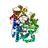
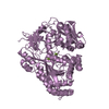
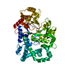
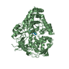
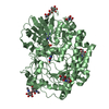
- PDB-2x1i: glycoside hydrolase family 77 4-alpha-glucanotransferase from the... -
+ Open data

- Basic information
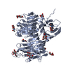
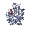
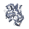
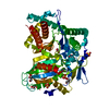
| Entry | Database: PDB / ID: 2x1i | ||||||
|---|---|---|---|---|---|---|---|
| Title | glycoside hydrolase family 77 4-alpha-glucanotransferase from thermus brockianus | ||||||
 Components Components | 4-ALPHA-GLUCANOTRANSFERASE | ||||||
 Keywords Keywords | TRANSFERASE | ||||||
| Function / homology |  Function and homology information Function and homology information4-alpha-glucanotransferase / 4-alpha-glucanotransferase activity / carbohydrate metabolic process Similarity search - Function | ||||||
| Biological species |   THERMUS BROCKIANUS (bacteria) THERMUS BROCKIANUS (bacteria) | ||||||
| Method |  X-RAY DIFFRACTION / SYNCHROTRON / MOLECULAR REPLACEMENT / Resolution: 2.36 Å X-RAY DIFFRACTION / SYNCHROTRON / MOLECULAR REPLACEMENT / Resolution: 2.36 Å | ||||||
 Authors Authors | Yoon, S.-M. / Jung, J.-H. / Jung, T.-Y. / Song, H.-N. / Park, C.-S. / Woo, E.-J. | ||||||
 Citation Citation | Journal: Proteins / Year: 2011 Title: Structural and Functional Analysis of Substrate Recognition by the 250S Loop in Amylomaltase from Thermus Brockianus. Authors: Jung, J.-H. / Jung, T.-Y. / Seo, D. / Yoon, S.-M. / Choi, H. / Park, B.C. / Park, C.-S. / Woo, E.-J. | ||||||
| History |
|
- Structure visualization
| Structure viewer | Molecule: MolmilJmol/JSmol |
|---|
- Downloads & links
-Download
| PDBx/mmCIF format | 2x1i.cif.gz | 114.4 KB | Display | PDBx/mmCIF format |
|---|---|---|---|---|
| PDB format | pdb2x1i.ent.gz | 88.1 KB | Display | PDB format |
| PDBx/mmJSON format | 2x1i.json.gz | Tree view | PDBx/mmJSON format | |
| Others |  Other downloads Other downloads |
-Validation report
| Arichive directory | https://data.pdbj.org/pub/pdb/validation_reports/x1/2x1iftp://data.pdbj.org/pub/pdb/validation_reports/x1/2x1i | HTTPS FTP |
|---|
-Related structure data
| Related structure data | 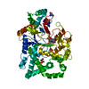 2owcS S: Starting model for refinement |
|---|---|
| Similar structure data |

-Links
 PDBj
PDBj- Assembly
| Deposited unit | 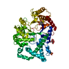
| ||||||||
|---|---|---|---|---|---|---|---|---|---|
| 1 |
| ||||||||
| Unit cell |
|
-Components
| #1: Protein | Mass: 57353.812 Da / Num. of mol.: 1 / Source method: isolated from a natural source / Source: (natural) THERMUS BROCKIANUS (bacteria) / Strain: JCM11602 / References: UniProt: Q2VJA0 | ||
|---|---|---|---|
| #2: Chemical | ChemComp-PO4 /   Mass: 94.971 Da / Num. of mol.: 1 / Source method: obtained synthetically / Formula: PO4 Mass: 94.971 Da / Num. of mol.: 1 / Source method: obtained synthetically / Formula: PO4 | ||
| #3: Chemical | ChemComp-SO4 /   Mass: 96.063 Da / Num. of mol.: 1 / Source method: obtained synthetically / Formula: SO4 Mass: 96.063 Da / Num. of mol.: 1 / Source method: obtained synthetically / Formula: SO4 | ||
| #4: Chemical |   Mass: 180.156 Da / Num. of mol.: 2 / Source method: obtained synthetically / Formula: C6H12O6 Mass: 180.156 Da / Num. of mol.: 2 / Source method: obtained synthetically / Formula: C6H12O6#5: Water | ChemComp-HOH / |  Mass: 18.015 Da / Num. of mol.: 110 / Source method: isolated from a natural source / Formula: H2O Mass: 18.015 Da / Num. of mol.: 110 / Source method: isolated from a natural source / Formula: H2O |
-Experimental details
-Experiment
| Experiment | Method: X-RAY DIFFRACTION / Number of used crystals: 1 |
|---|
- Sample preparation
| Crystal | Density Matthews: 3.57 Å3/Da / Density % sol: 65.61 % / Description: NONE |
|---|---|
| Crystal grow | pH: 6.5 Details: 0.1M BIS-TRIS PH6.5, 45% PPGP400, 12% INOSITOL(ADDITIVE) |
-Data collection
| Diffraction | Mean temperature: 294 K |
|---|---|
| Diffraction source | Source: SYNCHROTRON / Site: PAL/PLS  / Beamline: 4A / Wavelength: 1.2399 / Beamline: 4A / Wavelength: 1.2399 |
| Detector | Type: ADSC CCD / Detector: CCD / Date: Dec 5, 2008 |
| Radiation | Protocol: SINGLE WAVELENGTH / Monochromatic (M) / Laue (L): M / Scattering type: x-ray |
| Radiation wavelength | Wavelength: 1.2399 Å / Relative weight: 1 |
| Reflection | Resolution: 2.36→29.93 Å / Num. obs: 35592 / % possible obs: 99.8 % / Observed criterion σ(I): 0 / Redundancy: 21.4 % / Biso Wilson estimate: 21.1 Å2 / Rmerge(I) obs: 0.09 / Net I/σ(I): 34.06 |
| Reflection shell | Resolution: 2.36→2.44 Å / Redundancy: 14.6 % / Rmerge(I) obs: 0.36 / Mean I/σ(I) obs: 5.36 / % possible all: 96 |
- Processing
| Software |
| ||||||||||||||||||||||||||||||||||||||||||||||||||||||||||||||||||||||||||||||||
|---|---|---|---|---|---|---|---|---|---|---|---|---|---|---|---|---|---|---|---|---|---|---|---|---|---|---|---|---|---|---|---|---|---|---|---|---|---|---|---|---|---|---|---|---|---|---|---|---|---|---|---|---|---|---|---|---|---|---|---|---|---|---|---|---|---|---|---|---|---|---|---|---|---|---|---|---|---|---|---|---|---|
| Refinement | Method to determine structure: MOLECULAR REPLACEMENT Starting model: PDB ENTRY 2OWC Resolution: 2.36→29.93 Å / Rfactor Rfree error: 0.006 / Data cutoff high absF: 76946.94 / Data cutoff low absF: 0 / Isotropic thermal model: RESTRAINED / Cross valid method: THROUGHOUT / σ(F): 0 / Stereochemistry target values: MAXIMUM LIKELIHOOD / Details: BULK SOLVENT MODEL USED
| ||||||||||||||||||||||||||||||||||||||||||||||||||||||||||||||||||||||||||||||||
| Solvent computation | Solvent model: FLAT MODEL / Bsol: 33.5482 Å2 / ksol: 0.35 e/Å3 | ||||||||||||||||||||||||||||||||||||||||||||||||||||||||||||||||||||||||||||||||
| Displacement parameters | Biso mean: 39.1 Å2
| ||||||||||||||||||||||||||||||||||||||||||||||||||||||||||||||||||||||||||||||||
| Refine analyze |
| ||||||||||||||||||||||||||||||||||||||||||||||||||||||||||||||||||||||||||||||||
| Refinement step | Cycle: LAST / Resolution: 2.36→29.93 Å
| ||||||||||||||||||||||||||||||||||||||||||||||||||||||||||||||||||||||||||||||||
| Refine LS restraints |
| ||||||||||||||||||||||||||||||||||||||||||||||||||||||||||||||||||||||||||||||||
| Refine LS restraints NCS | NCS model details: NONE | ||||||||||||||||||||||||||||||||||||||||||||||||||||||||||||||||||||||||||||||||
| LS refinement shell | Resolution: 2.36→2.51 Å / Rfactor Rfree error: 0.069 / Total num. of bins used: 6
| ||||||||||||||||||||||||||||||||||||||||||||||||||||||||||||||||||||||||||||||||
| Xplor file |
|
