- PDB-3p3l: Crystal Structure of the Cytochrome P450 monooxygenase AurH (wild... -
+
Open data
ID or keywords:
Loading...
-
Basic information
Entry
Database: PDB / ID: 3p3l
Title
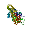
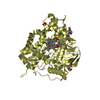
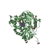
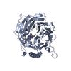
Crystal Structure of the Cytochrome P450 monooxygenase AurH (wildtype) from Streptomyces Thioluteus
Components
Cytochrome P450
Keywords
OXIDOREDUCTASE / Cytochrome P450 / Monooxygenase
Function / homology
Function and homology information
luteothin monooxygenase / oxidoreductase activity, acting on paired donors, with incorporation or reduction of molecular oxygen / antibiotic biosynthetic process / monooxygenase activity / iron ion binding / heme binding Similarity search - Function
Resolution: 2.05→2.1 Å / Redundancy: 2.7 % / Rmerge(I) obs: 0.615 / Mean I/σ(I) obs: 2.7 / Num. unique all: 4164 / Rsym value: 0.499 / % possible all: 99.6
-
Processing
Software
Name
Version
Classification
MAR345dtb
datacollection
PHASER
phasing
REFMAC
5.6.0085
refinement
XDS
datareduction
XDS
datascaling
Refinement
Method to determine structure: MOLECULAR REPLACEMENT / Resolution: 2.1→20 Å / Cor.coef. Fo:Fc: 0.919 / Cor.coef. Fo:Fc free: 0.874 / SU B: 11.783 / SU ML: 0.149 / Isotropic thermal model: TLS / Cross valid method: THROUGHOUT / ESU R Free: 0.208 / Stereochemistry target values: MAXIMUM LIKELIHOOD / Details: HYDROGENS HAVE BEEN USED IF PRESENT IN THE INPUT
Rfactor
Num. reflection
% reflection
Selection details
Rfree
0.26366
2606
5 %
RANDOM
Rwork
0.21448
-
-
-
obs
0.21693
49526
100 %
-
Solvent computation
Ion probe radii: 0.8 Å / Shrinkage radii: 0.8 Å / VDW probe radii: 1.2 Å / Solvent model: BABINET MODEL WITH MASK
Displacement parameters
Biso mean: 16.482 Å2
Baniso -1
Baniso -2
Baniso -3
1-
3.29 Å2
-0 Å2
1.62 Å2
2-
-
-1.09 Å2
0 Å2
3-
-
-
-0.42 Å2
Refinement step
Cycle: LAST / Resolution: 2.1→20 Å
Protein
Nucleic acid
Ligand
Solvent
Total
Num. atoms
6300
0
132
337
6769
Refine LS restraints
Refine-ID
Type
Dev ideal
Dev ideal target
Number
X-RAY DIFFRACTION
r_bond_refined_d
0.014
0.022
6612
X-RAY DIFFRACTION
r_bond_other_d
0.002
0.02
4418
X-RAY DIFFRACTION
r_angle_refined_deg
0.735
2.005
9062
X-RAY DIFFRACTION
r_angle_other_deg
0.688
3
10718
X-RAY DIFFRACTION
r_dihedral_angle_1_deg
5.961
5
818
X-RAY DIFFRACTION
r_dihedral_angle_2_deg
33.853
23.377
308
X-RAY DIFFRACTION
r_dihedral_angle_3_deg
14.121
15
1014
X-RAY DIFFRACTION
r_dihedral_angle_4_deg
12.695
15
56
X-RAY DIFFRACTION
r_chiral_restr
0.06
0.2
1000
X-RAY DIFFRACTION
r_gen_planes_refined
0.005
0.021
7433
X-RAY DIFFRACTION
r_gen_planes_other
0.001
0.02
1355
LS refinement shell
Resolution: 2.1→2.154 Å / Total num. of bins used: 20
Rfactor
Num. reflection
% reflection
Rfree
0.257
188
-
Rwork
0.24
3586
-
obs
-
-
100 %
Refinement TLS params.
Method: refined / Refine-ID: X-RAY DIFFRACTION
ID
L11 (°2)
L12 (°2)
L13 (°2)
L22 (°2)
L23 (°2)
L33 (°2)
S11 (Å °)
S12 (Å °)
S13 (Å °)
S21 (Å °)
S22 (Å °)
S23 (Å °)
S31 (Å °)
S32 (Å °)
S33 (Å °)
T11 (Å2)
T12 (Å2)
T13 (Å2)
T22 (Å2)
T23 (Å2)
T33 (Å2)
Origin x (Å)
Origin y (Å)
Origin z (Å)
1
0.1067
0.008
0.0386
0.3304
0.2115
0.2099
0.029
0.0112
-0.0158
-0.0143
-0.0699
0.0395
-0.0572
-0.039
0.0409
0.0792
0.0122
-0.0055
0.0595
-0.0095
0.0315
-3.206
12.603
37.6211
2
0.1514
0.0502
-0.0266
0.287
0.0183
0.2125
0.0086
-0.0024
-0.0216
0.0124
-0.0252
-0.019
-0.0016
-0.0047
0.0166
0.0072
-0.0012
-0.0205
0.0627
0.004
0.0791
-20.5606
34.9633
-1.7313
3
0.0019
-0.0005
-0.0031
0.0004
-0.001
0.0186
-0.0092
-0.0197
0.0048
-0.0006
-0.0067
-0.0053
0.0252
0.0951
0.0159
0.0356
0.1326
0.0175
0.4956
0.076
0.0689
-21.3031
29.7833
6.1182
4
0.0394
-0.0253
0.0481
0.0659
-0.033
0.067
-0.0014
0.012
0.0096
0.0198
-0.0194
0.0191
0.0012
0.009
0.0208
0.0306
-0.0092
-0.0104
0.0764
-0.0069
0.0787
-11.3492
25.4508
15.7077
Refinement TLS group
ID
Refine-ID
Refine TLS-ID
Auth asym-ID
Auth seq-ID
1
X-RAY DIFFRACTION
1
A
4 - 406
2
X-RAY DIFFRACTION
1
A
501
3
X-RAY DIFFRACTION
2
B
4 - 406
4
X-RAY DIFFRACTION
2
B
501
5
X-RAY DIFFRACTION
3
A
407 - 409
6
X-RAY DIFFRACTION
3
B
407 - 409
7
X-RAY DIFFRACTION
4
A
410
8
X-RAY DIFFRACTION
4
B
410 - 745
+
About Yorodumi
-
News
-
Feb 9, 2022. New format data for meta-information of EMDB entries
New format data for meta-information of EMDB entries
Version 3 of the EMDB header file is now the official format.
The previous official version 1.9 will be removed from the archive.
In the structure databanks used in Yorodumi, some data are registered as the other names, "COVID-19 virus" and "2019-nCoV". Here are the details of the virus and the list of structure data.
Jan 31, 2019. EMDB accession codes are about to change! (news from PDBe EMDB page)
EMDB accession codes are about to change! (news from PDBe EMDB page)
The allocation of 4 digits for EMDB accession codes will soon come to an end. Whilst these codes will remain in use, new EMDB accession codes will include an additional digit and will expand incrementally as the available range of codes is exhausted. The current 4-digit format prefixed with “EMD-” (i.e. EMD-XXXX) will advance to a 5-digit format (i.e. EMD-XXXXX), and so on. It is currently estimated that the 4-digit codes will be depleted around Spring 2019, at which point the 5-digit format will come into force.
The EM Navigator/Yorodumi systems omit the EMD- prefix.
Related info.:Q: What is EMD? / ID/Accession-code notation in Yorodumi/EM Navigator
Yorodumi is a browser for structure data from EMDB, PDB, SASBDB, etc.
This page is also the successor to EM Navigator detail page, and also detail information page/front-end page for Omokage search.
The word "yorodu" (or yorozu) is an old Japanese word meaning "ten thousand". "mi" (miru) is to see.
Related info.:EMDB / PDB / SASBDB / Comparison of 3 databanks / Yorodumi Search / Aug 31, 2016. New EM Navigator & Yorodumi / Yorodumi Papers / Jmol/JSmol / Function and homology information / Changes in new EM Navigator and Yorodumi
 Movie
Movie Controller

 Yorodumi
 Open data
 Basic information
 Components
 Keywords
 Function and homology information
 Streptomyces thioluteus (bacteria)
 X-RAY DIFFRACTION /
  Authors
 Citation
 Structure visualization
 Downloads & links
 Other downloads


 PDBj
PDBj
 Assembly


 Mass: 616.487 Da / Num. of mol.: 2 / Source method: obtained synthetically / Formula: C34H32FeN4O4
 Mass: 96.063 Da / Num. of mol.: 3 / Source method: obtained synthetically / Formula: SO4
 Mass: 238.305 Da / Num. of mol.: 2 / Source method: obtained synthetically / Formula: C8H18N2O4S / Comment: pH buffer*YM
 Mass: 35.453 Da / Num. of mol.: 1 / Source method: obtained synthetically / Formula: Cl
 Sample preparation
 / Beamline: 14.2 / Wavelength: 0.91841 Å
 Processing