- PDB-3p3x: Crystal Structure of the Cytochrome P450 Monooxygenase AurH (nter... -
+
Open data
ID or keywords:
Loading...
-
Basic information
Entry
Database: PDB / ID: 3p3x
Title
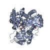
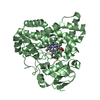
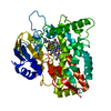
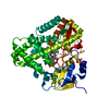
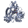
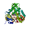
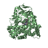
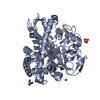
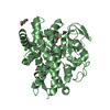
Crystal Structure of the Cytochrome P450 Monooxygenase AurH (nterm-AurH-I) from Streptomyces Thioluteus
Components
Cytochrome P450
Keywords
OXIDOREDUCTASE / Cytochrome P450 Monooxygenase / Oxidation of Deoxyaureothin to Aureothin
Function / homology
Function and homology information
luteothin monooxygenase / oxidoreductase activity, acting on paired donors, with incorporation or reduction of molecular oxygen / antibiotic biosynthetic process / monooxygenase activity / iron ion binding / heme binding Similarity search - Function
Method to determine structure: MOLECULAR REPLACEMENT / Resolution: 2.3→30 Å / Cor.coef. Fo:Fc: 0.931 / Cor.coef. Fo:Fc free: 0.902 / SU B: 20.765 / SU ML: 0.233 / Cross valid method: THROUGHOUT / ESU R Free: 0.276 / Stereochemistry target values: MAXIMUM LIKELIHOOD / Details: HYDROGENS HAVE BEEN ADDED IN THE RIDING POSITIONS
Rfactor
Num. reflection
% reflection
Selection details
Rfree
0.27957
1525
4 %
RANDOM
Rwork
0.22562
-
-
-
obs
0.22783
36599
100 %
-
all
-
55540
-
-
Solvent computation
Ion probe radii: 0.8 Å / Shrinkage radii: 0.8 Å / VDW probe radii: 1.4 Å / Solvent model: BABINET MODEL WITH MASK
Displacement parameters
Biso mean: 11.596 Å2
Baniso -1
Baniso -2
Baniso -3
1-
4.42 Å2
-0 Å2
-0 Å2
2-
-
-3.01 Å2
0 Å2
3-
-
-
-1.42 Å2
Refinement step
Cycle: LAST / Resolution: 2.3→30 Å
Protein
Nucleic acid
Ligand
Solvent
Total
Num. atoms
6239
0
111
208
6558
Refine LS restraints
Refine-ID
Type
Dev ideal
Dev ideal target
Number
X-RAY DIFFRACTION
r_bond_refined_d
0.011
0.022
6521
X-RAY DIFFRACTION
r_bond_other_d
X-RAY DIFFRACTION
r_angle_refined_deg
1.319
2.008
8922
X-RAY DIFFRACTION
r_angle_other_deg
X-RAY DIFFRACTION
r_dihedral_angle_1_deg
5.626
5
794
X-RAY DIFFRACTION
r_dihedral_angle_2_deg
35.885
23.344
305
X-RAY DIFFRACTION
r_dihedral_angle_3_deg
18.141
15
996
X-RAY DIFFRACTION
r_dihedral_angle_4_deg
14.339
15
56
X-RAY DIFFRACTION
r_chiral_restr
0.09
0.2
983
X-RAY DIFFRACTION
r_gen_planes_refined
0.006
0.021
5070
X-RAY DIFFRACTION
r_gen_planes_other
X-RAY DIFFRACTION
r_nbd_refined
X-RAY DIFFRACTION
r_nbd_other
X-RAY DIFFRACTION
r_nbtor_refined
X-RAY DIFFRACTION
r_nbtor_other
X-RAY DIFFRACTION
r_xyhbond_nbd_refined
X-RAY DIFFRACTION
r_xyhbond_nbd_other
X-RAY DIFFRACTION
r_metal_ion_refined
X-RAY DIFFRACTION
r_metal_ion_other
X-RAY DIFFRACTION
r_symmetry_vdw_refined
X-RAY DIFFRACTION
r_symmetry_vdw_other
X-RAY DIFFRACTION
r_symmetry_hbond_refined
X-RAY DIFFRACTION
r_symmetry_hbond_other
X-RAY DIFFRACTION
r_symmetry_metal_ion_refined
X-RAY DIFFRACTION
r_symmetry_metal_ion_other
X-RAY DIFFRACTION
r_mcbond_it
1.846
5
3988
X-RAY DIFFRACTION
r_mcbond_other
X-RAY DIFFRACTION
r_mcangle_it
2.48
6
6445
X-RAY DIFFRACTION
r_scbond_it
2.987
7
2533
X-RAY DIFFRACTION
r_scangle_it
3.38
7
2477
X-RAY DIFFRACTION
r_rigid_bond_restr
X-RAY DIFFRACTION
r_sphericity_free
X-RAY DIFFRACTION
r_sphericity_bonded
LS refinement shell
Resolution: 2.3→2.359 Å / Total num. of bins used: 20
Rfactor
Num. reflection
% reflection
Rfree
0.308
108
-
Rwork
0.264
2579
-
obs
-
-
100 %
Refinement TLS params.
Method: refined / Refine-ID: X-RAY DIFFRACTION
ID
L11 (°2)
L12 (°2)
L13 (°2)
L22 (°2)
L23 (°2)
L33 (°2)
S11 (Å °)
S12 (Å °)
S13 (Å °)
S21 (Å °)
S22 (Å °)
S23 (Å °)
S31 (Å °)
S32 (Å °)
S33 (Å °)
T11 (Å2)
T12 (Å2)
T13 (Å2)
T22 (Å2)
T23 (Å2)
T33 (Å2)
Origin x (Å)
Origin y (Å)
Origin z (Å)
1
0.5674
-0.1847
-0.2847
1.0623
-0.3939
1.6137
0.002
0.0607
-0.0312
-0.0383
0.0821
0.1476
0.0878
-0.0376
-0.0841
0.0563
-0.0235
-0.011
0.1252
0.0045
0.1718
18.7905
10.1398
23.9032
2
0.7964
-0.0879
-0.0009
0.9047
0.8047
2.3175
0.0353
0.0627
0.0238
-0.0564
0.1265
-0.201
0.0741
0.1744
-0.1619
0.1088
-0.0676
0.0117
0.1587
-0.025
0.1784
46.6418
32.997
50.4296
3
0.9208
0.456
1.1086
0.2426
0.5774
1.4146
-0.3781
0.3525
-0.0582
-0.1715
0.2933
-0.04
-0.4169
0.5676
0.0848
0.5995
-0.091
0.117
0.4719
0.0916
0.4259
36.1691
18.0642
27.9632
4
0.1285
-0.0212
0.078
0.2643
0.3083
0.4963
0.0169
0.0161
-0.0129
-0.0278
0.0249
-0.02
-0.0294
0.0243
-0.0419
0.0085
-0.0215
0.0036
0.2409
0.0183
0.2028
30.7139
21.483
36.4848
Refinement TLS group
ID
Refine-ID
Refine TLS-ID
Auth asym-ID
Auth seq-ID
1
X-RAY DIFFRACTION
1
A
8 - 404
2
X-RAY DIFFRACTION
1
A
501
3
X-RAY DIFFRACTION
2
B
6 - 404
4
X-RAY DIFFRACTION
2
B
501
5
X-RAY DIFFRACTION
3
A
407 - 409
6
X-RAY DIFFRACTION
3
B
407 - 409
7
X-RAY DIFFRACTION
4
A
413 - 525
8
X-RAY DIFFRACTION
4
B
410 - 503
+
About Yorodumi
-
News
-
Feb 9, 2022. New format data for meta-information of EMDB entries
New format data for meta-information of EMDB entries
Version 3 of the EMDB header file is now the official format.
The previous official version 1.9 will be removed from the archive.
In the structure databanks used in Yorodumi, some data are registered as the other names, "COVID-19 virus" and "2019-nCoV". Here are the details of the virus and the list of structure data.
Jan 31, 2019. EMDB accession codes are about to change! (news from PDBe EMDB page)
EMDB accession codes are about to change! (news from PDBe EMDB page)
The allocation of 4 digits for EMDB accession codes will soon come to an end. Whilst these codes will remain in use, new EMDB accession codes will include an additional digit and will expand incrementally as the available range of codes is exhausted. The current 4-digit format prefixed with “EMD-” (i.e. EMD-XXXX) will advance to a 5-digit format (i.e. EMD-XXXXX), and so on. It is currently estimated that the 4-digit codes will be depleted around Spring 2019, at which point the 5-digit format will come into force.
The EM Navigator/Yorodumi systems omit the EMD- prefix.
Related info.:Q: What is EMD? / ID/Accession-code notation in Yorodumi/EM Navigator
Yorodumi is a browser for structure data from EMDB, PDB, SASBDB, etc.
This page is also the successor to EM Navigator detail page, and also detail information page/front-end page for Omokage search.
The word "yorodu" (or yorozu) is an old Japanese word meaning "ten thousand". "mi" (miru) is to see.
Related info.:EMDB / PDB / SASBDB / Comparison of 3 databanks / Yorodumi Search / Aug 31, 2016. New EM Navigator & Yorodumi / Yorodumi Papers / Jmol/JSmol / Function and homology information / Changes in new EM Navigator and Yorodumi
 Movie
Movie Controller

 Yorodumi
 Open data
 Basic information
 Components
 Keywords
 Function and homology information
 Streptomyces Thioluteus (bacteria)
 X-RAY DIFFRACTION /
  Authors
 Citation
 Structure visualization
 Downloads & links
 Other downloads


 PDBj
PDBj
 Assembly


 Mass: 616.487 Da / Num. of mol.: 2 / Source method: obtained synthetically / Formula: C34H32FeN4O4
 Mass: 92.094 Da / Num. of mol.: 3 / Source method: obtained synthetically / Formula: C3H8O3
 Mass: 96.063 Da / Num. of mol.: 1 / Source method: obtained synthetically / Formula: SO4
 Mass: 35.453 Da / Num. of mol.: 2 / Source method: obtained synthetically / Formula: Cl
 Sample preparation
 / Beamline: 14.2 / Wavelength: 0.91841 Å
 Processing