 Movie
Movie Controller

[English] 日本語
 Yorodumi
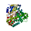
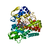
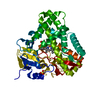
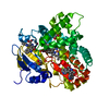
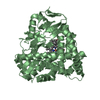
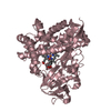
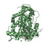
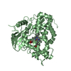
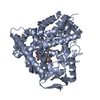
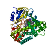
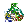
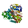
- PDB-2z3t: Crystal Structure of Substrate Free Cytochrome P450 StaP (CYP245A1) -
+ Open data

- Basic information
| Entry | Database: PDB / ID: 2z3t | ||||||
|---|---|---|---|---|---|---|---|
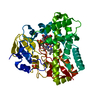
| Title | Crystal Structure of Substrate Free Cytochrome P450 StaP (CYP245A1) | ||||||
 Components Components | Cytochrome P450 | ||||||
 Keywords Keywords | OXIDOREDUCTASE / cytochrome P450 / monoxygenase / oxydoreductase / heme-enzyme | ||||||
| Function / homology |  Function and homology information Function and homology informationoxidoreductase activity, acting on paired donors, with incorporation or reduction of molecular oxygen / monooxygenase activity / iron ion binding / heme binding Similarity search - Function | ||||||
| Biological species |  Streptomyces sp. TP-A0274 (bacteria) Streptomyces sp. TP-A0274 (bacteria) | ||||||
| Method |  X-RAY DIFFRACTION / SYNCHROTRON / MAD / Resolution: 1.9 Å X-RAY DIFFRACTION / SYNCHROTRON / MAD / Resolution: 1.9 Å | ||||||
 Authors Authors | Makino, M. / Sugimoto, H. / Shiro, Y. / Asamizu, S. / Onaka, H. / Nagano, S. | ||||||
 Citation Citation | Journal: Proc.Natl.Acad.Sci.Usa / Year: 2007 Title: Crystal structures and catalytic mechanism of cytochrome P450 StaP that produces the indolocarbazole skeleton Authors: Makino, M. / Sugimoto, H. / Shiro, Y. / Asamizu, S. / Onaka, H. / Nagano, S. | ||||||
| History |
|
- Structure visualization
| Structure viewer | Molecule: MolmilJmol/JSmol |
|---|
- Downloads & links
-Download
| PDBx/mmCIF format | 2z3t.cif.gz | 318.2 KB | Display | PDBx/mmCIF format |
|---|---|---|---|---|
| PDB format | pdb2z3t.ent.gz | 259 KB | Display | PDB format |
| PDBx/mmJSON format | 2z3t.json.gz | Tree view | PDBx/mmJSON format | |
| Others |  Other downloads Other downloads |
-Validation report
| Arichive directory | https://data.pdbj.org/pub/pdb/validation_reports/z3/2z3tftp://data.pdbj.org/pub/pdb/validation_reports/z3/2z3t | HTTPS FTP |
|---|
-Related structure data


-Links
 PDBj
PDBj


- Assembly
| Deposited unit | 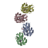
| ||||||||
|---|---|---|---|---|---|---|---|---|---|
| 1 | 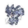
| ||||||||
| 2 | 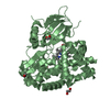
| ||||||||
| 3 | 
| ||||||||
| 4 | 
| ||||||||
| Unit cell |
| ||||||||
| Details | The biological assembly is a monomer. |
-Components
| #1: Protein | Mass: 47262.262 Da / Num. of mol.: 4 Source method: isolated from a genetically manipulated source Source: (gene. exp.) Streptomyces sp. TP-A0274 (bacteria) / Gene: staP / Plasmid: pET26b / Species (production host): Escherichia coli / Production host: #2: Chemical | ChemComp-HEM /   Mass: 616.487 Da / Num. of mol.: 4 / Source method: obtained synthetically / Formula: C34H32FeN4O4 Mass: 616.487 Da / Num. of mol.: 4 / Source method: obtained synthetically / Formula: C34H32FeN4O4#3: Chemical | ChemComp-IMD /   Mass: 69.085 Da / Num. of mol.: 4 / Source method: obtained synthetically / Formula: C3H5N2 Mass: 69.085 Da / Num. of mol.: 4 / Source method: obtained synthetically / Formula: C3H5N2#4: Chemical | ChemComp-EDO /   Mass: 62.068 Da / Num. of mol.: 9 / Source method: obtained synthetically / Formula: C2H6O2 Mass: 62.068 Da / Num. of mol.: 9 / Source method: obtained synthetically / Formula: C2H6O2#5: Water | ChemComp-HOH / |  Mass: 18.015 Da / Num. of mol.: 426 / Source method: isolated from a natural source / Formula: H2O Mass: 18.015 Da / Num. of mol.: 426 / Source method: isolated from a natural source / Formula: H2O |
|---|
-Experimental details
-Experiment
| Experiment | Method: X-RAY DIFFRACTION / Number of used crystals: 1 |
|---|
- Sample preparation
| Crystal | Density Matthews: 2.58 Å3/Da / Density % sol: 52.41 % |
|---|---|
| Crystal grow | Temperature: 293 K / Method: vapor diffusion / pH: 8 Details: 22% PEG 3350, 50mM potassium fluoride, 100mM imidazole, pH 8.0, VAPOR DIFFUSION, temperature 293K |
-Data collection
| Diffraction |
| ||||||||||||||||||
|---|---|---|---|---|---|---|---|---|---|---|---|---|---|---|---|---|---|---|---|
| Diffraction source |
| ||||||||||||||||||
| Detector |
| ||||||||||||||||||
| Radiation |
| ||||||||||||||||||
| Radiation wavelength |
| ||||||||||||||||||
| Reflection twin | Type: hemihedral / Operator: -h,-k,h+l / Fraction: 0.453 | ||||||||||||||||||
| Reflection | Resolution: 1.9→20 Å / Num. obs: 138934 / % possible obs: 91.4 % / Observed criterion σ(I): 0 / Redundancy: 3.3 % / Biso Wilson estimate: 31.42 Å2 / Rsym value: 0.032 / Net I/σ(I): 20.66 | ||||||||||||||||||
| Reflection shell | Resolution: 1.9→1.97 Å / Redundancy: 3.2 % / Mean I/σ(I) obs: 3.34 / Num. unique all: 10180 / Rsym value: 0.416 / % possible all: 67.1 |

- Processing
| Software |
| ||||||||||||||||||||||||||||
|---|---|---|---|---|---|---|---|---|---|---|---|---|---|---|---|---|---|---|---|---|---|---|---|---|---|---|---|---|---|
| Refinement | Method to determine structure: MAD / Resolution: 1.9→20 Å / Cross valid method: THROUGHOUT / σ(F): 11960 Details: Used twin_lsq target. Twin fraction 0.453. Twin low -h, -k, h+l
| ||||||||||||||||||||||||||||
| Solvent computation | Bsol: 59.704 Å2 | ||||||||||||||||||||||||||||
| Displacement parameters | Biso mean: 43.856 Å2
| ||||||||||||||||||||||||||||
| Refine analyze |
| ||||||||||||||||||||||||||||
| Refinement step | Cycle: LAST / Resolution: 1.9→20 Å
| ||||||||||||||||||||||||||||
| Refine LS restraints |
| ||||||||||||||||||||||||||||
| LS refinement shell | Resolution: 1.9→1.97 Å
| ||||||||||||||||||||||||||||
| Xplor file |
|
