 Movie
Movie Controller

[English] 日本語
 Yorodumi
- PDB-2z3u: Crystal Structure of Chromopyrrolic Acid Bound Cytochrome P450 St... -
+ Open data

- Basic information
| Entry | Database: PDB / ID: 2z3u | ||||||
|---|---|---|---|---|---|---|---|
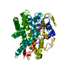
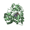
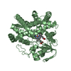
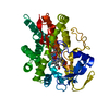
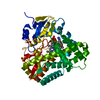
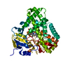
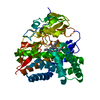
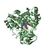
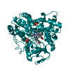
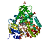
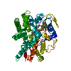
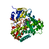
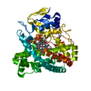
| Title | Crystal Structure of Chromopyrrolic Acid Bound Cytochrome P450 StaP (CYP245A1) | ||||||
 Components Components | Cytochrome P450 | ||||||
 Keywords Keywords | OXIDOREDUCTASE / cytochrome P450 / monoxygenase / oxydoreductase / heme-enzyme / chromopyrrolic acid | ||||||
| Function / homology |  Function and homology information Function and homology informationoxidoreductase activity, acting on paired donors, with incorporation or reduction of molecular oxygen / monooxygenase activity / iron ion binding / heme binding Similarity search - Function | ||||||
| Biological species |  Streptomyces sp. TP-A0274 (bacteria) Streptomyces sp. TP-A0274 (bacteria) | ||||||
| Method |  X-RAY DIFFRACTION / SYNCHROTRON / MOLECULAR REPLACEMENT / Resolution: 2.4 Å X-RAY DIFFRACTION / SYNCHROTRON / MOLECULAR REPLACEMENT / Resolution: 2.4 Å | ||||||
 Authors Authors | Makino, M. / Sugimoto, H. / Shiro, Y. / Asamizu, S. / Onaka, H. / Nagano, S. | ||||||
 Citation Citation | Journal: Proc.Natl.Acad.Sci.Usa / Year: 2007 Title: Crystal structures and catalytic mechanism of cytochrome P450 StaP that produces the indolocarbazole skeleton Authors: Makino, M. / Sugimoto, H. / Shiro, Y. / Asamizu, S. / Onaka, H. / Nagano, S. #1: Journal: J.Am.Chem.Soc. / Year: 2009Title: Theoretical and experimental studies of the conversion of chromopyrrolic acid to an antitumor derivative by cytochrome P450 StaP: the catalytic role of water molecules Authors: Wang, Y. / Chen, H. / Makino, M. / Shiro, Y. / Nagano, S. / Asamizu, S. / Onaka, H. / Shaik, S. | ||||||
| History |
|
- Structure visualization
| Structure viewer | Molecule: MolmilJmol/JSmol |
|---|
- Downloads & links
-Download
| PDBx/mmCIF format | 2z3u.cif.gz | 104.2 KB | Display | PDBx/mmCIF format |
|---|---|---|---|---|
| PDB format | pdb2z3u.ent.gz | 77.4 KB | Display | PDB format |
| PDBx/mmJSON format | 2z3u.json.gz | Tree view | PDBx/mmJSON format | |
| Others |  Other downloads Other downloads |
-Validation report
| Arichive directory | https://data.pdbj.org/pub/pdb/validation_reports/z3/2z3uftp://data.pdbj.org/pub/pdb/validation_reports/z3/2z3u | HTTPS FTP |
|---|
-Related structure data
| Related structure data | 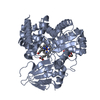 2z3tSC S: Starting model for refinement C: citing same article ( |
|---|---|
| Similar structure data |


-Links
 PDBj
PDBj
- Assembly
| Deposited unit | 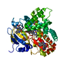
| ||||||||
|---|---|---|---|---|---|---|---|---|---|
| 1 |
| ||||||||
| Unit cell |
|
-Components
| #1: Protein | Mass: 47262.262 Da / Num. of mol.: 1 Source method: isolated from a genetically manipulated source Source: (gene. exp.) Streptomyces sp. TP-A0274 (bacteria) / Gene: staP / Plasmid: pET26b / Species (production host): Escherichia coli / Production host: | ||||
|---|---|---|---|---|---|
| #2: Chemical | ChemComp-HEM /   Mass: 616.487 Da / Num. of mol.: 1 / Source method: obtained synthetically / Formula: C34H32FeN4O4 Mass: 616.487 Da / Num. of mol.: 1 / Source method: obtained synthetically / Formula: C34H32FeN4O4 | ||||
| #3: Chemical |   Mass: 385.372 Da / Num. of mol.: 3 / Source method: obtained synthetically / Formula: C22H15N3O4 Mass: 385.372 Da / Num. of mol.: 3 / Source method: obtained synthetically / Formula: C22H15N3O4#4: Chemical | ChemComp-EDO /   Mass: 62.068 Da / Num. of mol.: 4 / Source method: obtained synthetically / Formula: C2H6O2 Mass: 62.068 Da / Num. of mol.: 4 / Source method: obtained synthetically / Formula: C2H6O2#5: Water | ChemComp-HOH / |  Mass: 18.015 Da / Num. of mol.: 188 / Source method: isolated from a natural source / Formula: H2O Mass: 18.015 Da / Num. of mol.: 188 / Source method: isolated from a natural source / Formula: H2O |
-Experimental details
-Experiment
| Experiment | Method: X-RAY DIFFRACTION / Number of used crystals: 1 |
|---|
- Sample preparation
| Crystal | Density Matthews: 2.15 Å3/Da / Density % sol: 42.79 % |
|---|---|
| Crystal grow | Temperature: 293 K / Method: vapor diffusion / pH: 6.8 Details: 2% PEG 4000, 50mM Bis-Tris pH 6.8, 100mM magnesium chloride, 10mM manganese chloride, 5mM chromopyrrolic acid, VAPOR DIFFUSION, temperature 293K |
-Data collection
| Diffraction | Mean temperature: 90 K |
|---|---|
| Diffraction source | Source: SYNCHROTRON / Site: SPring-8  / Beamline: BL44B2 / Wavelength: 1 Å / Beamline: BL44B2 / Wavelength: 1 Å |
| Detector | Type: ADSC QUANTUM 210 / Detector: CCD / Date: Dec 15, 2006 |
| Radiation | Monochromator: Si / Protocol: SINGLE WAVELENGTH / Monochromatic (M) / Laue (L): M / Scattering type: x-ray |
| Radiation wavelength | Wavelength: 1 Å / Relative weight: 1 |
| Reflection | Resolution: 2.4→50 Å / Num. obs: 16573 / % possible obs: 99.2 % / Observed criterion σ(I): 0 / Redundancy: 6.1 % / Biso Wilson estimate: 20.2 Å2 / Rsym value: 0.074 / Net I/σ(I): 22.46 |
| Reflection shell | Resolution: 2.4→2.49 Å / Redundancy: 5.6 % / Mean I/σ(I) obs: 6.06 / Num. unique all: 1626 / Rsym value: 0.324 / % possible all: 97.7 |
-Phasing
| Phasing MR |
|
|---|
- Processing
| Software |
| ||||||||||||||||||||||||||||
|---|---|---|---|---|---|---|---|---|---|---|---|---|---|---|---|---|---|---|---|---|---|---|---|---|---|---|---|---|---|
| Refinement | Method to determine structure: MOLECULAR REPLACEMENT Starting model: PDB ENTRY 2Z3T Resolution: 2.4→19.96 Å / Rfactor Rfree error: 0.008 / Data cutoff high absF: 1570890.875 / Data cutoff low absF: 0 / Isotropic thermal model: RESTRAINED / Cross valid method: THROUGHOUT / σ(F): 0
| ||||||||||||||||||||||||||||
| Solvent computation | Solvent model: FLAT MODEL / Bsol: 41.309 Å2 / ksol: 0.356 e/Å3 | ||||||||||||||||||||||||||||
| Displacement parameters | Biso mean: 28.6 Å2
| ||||||||||||||||||||||||||||
| Refine analyze |
| ||||||||||||||||||||||||||||
| Refinement step | Cycle: LAST / Resolution: 2.4→19.96 Å
| ||||||||||||||||||||||||||||
| Refine LS restraints |
| ||||||||||||||||||||||||||||
| LS refinement shell | Resolution: 2.4→2.55 Å / Rfactor Rfree error: 0.026 / Total num. of bins used: 6
| ||||||||||||||||||||||||||||
| Xplor file |
|
