 Movie
Movie Controller


+ Open data

- Basic information
| Entry | Database: PDB / ID: 6anv | |||||||||
|---|---|---|---|---|---|---|---|---|---|---|
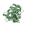
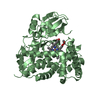
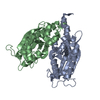
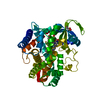
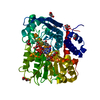
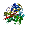
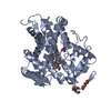
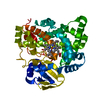
| Title | Crystal structure of anti-CRISPR protein AcrF1 | |||||||||
 Components Components | anti-CRISPR protein AcrF1 fused with C-terminal MBP tag | |||||||||
 Keywords Keywords | IMMUNE SYSTEM / Type I-F CRISPR-Cas system: Csy Cascade: Structure: anti-CRISPR protein: Inhibition of Csy complex: Genome editing tool | |||||||||
| Function / homology |  Function and homology information Function and homology informationcarbohydrate transmembrane transporter activity / maltose binding / maltose transport / maltodextrin transmembrane transport / ATP-binding cassette (ABC) transporter complex, substrate-binding subunit-containing / outer membrane-bounded periplasmic space Similarity search - Function | |||||||||
| Biological species |  Pseudomonas phage JBD30 (virus) Pseudomonas phage JBD30 (virus) | |||||||||
| Method |  X-RAY DIFFRACTION / SYNCHROTRON / MOLECULAR REPLACEMENT / molecular replacement / Resolution: 2.265 Å X-RAY DIFFRACTION / SYNCHROTRON / MOLECULAR REPLACEMENT / molecular replacement / Resolution: 2.265 Å | |||||||||
 Authors Authors | Yang, H. / Patel, D.J. | |||||||||
| Funding support |  United States, 2items United States, 2items
| |||||||||
 Citation Citation | Journal: Cell / Year: 2017 Title: Cryo-EM Structures Reveal Mechanism and Inhibition of DNA Targeting by a CRISPR-Cas Surveillance Complex. Authors: Tai Wei Guo / Alberto Bartesaghi / Hui Yang / Veronica Falconieri / Prashant Rao / Alan Merk / Edward T Eng / Ashleigh M Raczkowski / Tara Fox / Lesley A Earl / Dinshaw J Patel / Sriram Subramaniam / Abstract: Prokaryotic cells possess CRISPR-mediated adaptive immune systems that protect them from foreign genetic elements, such as invading viruses. A central element of this immune system is an RNA-guided ...Prokaryotic cells possess CRISPR-mediated adaptive immune systems that protect them from foreign genetic elements, such as invading viruses. A central element of this immune system is an RNA-guided surveillance complex capable of targeting non-self DNA or RNA for degradation in a sequence- and site-specific manner analogous to RNA interference. Although the complexes display considerable diversity in their composition and architecture, many basic mechanisms underlying target recognition and cleavage are highly conserved. Using cryoelectron microscopy (cryo-EM), we show that the binding of target double-stranded DNA (dsDNA) to a type I-F CRISPR system yersinia (Csy) surveillance complex leads to large quaternary and tertiary structural changes in the complex that are likely necessary in the pathway leading to target dsDNA degradation by a trans-acting helicase-nuclease. Comparison of the structure of the surveillance complex before and after dsDNA binding, or in complex with three virally encoded anti-CRISPR suppressors that inhibit dsDNA binding, reveals mechanistic details underlying target recognition and inhibition. | |||||||||
| History |
|
- Structure visualization
| Structure viewer | Molecule: MolmilJmol/JSmol |
|---|
- Downloads & links
-Download
| PDBx/mmCIF format | 6anv.cif.gz | 372.6 KB | Display | PDBx/mmCIF format |
|---|---|---|---|---|
| PDB format | pdb6anv.ent.gz | 304.1 KB | Display | PDB format |
| PDBx/mmJSON format | 6anv.json.gz | Tree view | PDBx/mmJSON format | |
| Others |  Other downloads Other downloads |
-Validation report
| Arichive directory | https://data.pdbj.org/pub/pdb/validation_reports/an/6anvftp://data.pdbj.org/pub/pdb/validation_reports/an/6anv | HTTPS FTP |
|---|
-Related structure data
| Related structure data | 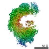 7048C  7049C  7050C  7051C  7052C 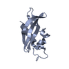 6anwC 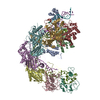 6b44C  6b45C 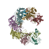 6b46C  6b47C  6b48C  4exkS S: Starting model for refinement C: citing same article ( |
|---|---|
| Similar structure data |
-Links
 PDBj
PDBj


- Assembly
| Deposited unit | 
| ||||||||
|---|---|---|---|---|---|---|---|---|---|
| 1 | 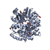
| ||||||||
| 2 | 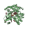
| ||||||||
| Unit cell |
|
-Components
-Protein / Sugars , 2 types, 4 molecules AB
| #1: Protein | Mass: 49773.141 Da / Num. of mol.: 2 Source method: isolated from a genetically manipulated source Source: (gene. exp.) Pseudomonas phage JBD30 (virus), (gene. exp.) Gene: JBD30_035, malE, Z5632, ECs5017 / Plasmid: pRSFDuet-SUMO / Production host: #2: Polysaccharide | 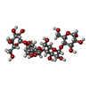  Source method: isolated from a genetically manipulated source Details: oligosaccharide / References: alpha-maltotetraose |
|---|
-Non-polymers , 5 types, 272 molecules 


| #3: Chemical |  Mass: 195.237 Da / Num. of mol.: 2 / Source method: obtained synthetically / Formula: C6H13NO4S / Comment: pH buffer*YM Mass: 195.237 Da / Num. of mol.: 2 / Source method: obtained synthetically / Formula: C6H13NO4S / Comment: pH buffer*YM#4: Chemical |  Mass: 150.173 Da / Num. of mol.: 3 / Source method: obtained synthetically / Formula: C6H14O4 Mass: 150.173 Da / Num. of mol.: 3 / Source method: obtained synthetically / Formula: C6H14O4#5: Chemical | ChemComp-EDO /  Mass: 62.068 Da / Num. of mol.: 7 / Source method: obtained synthetically / Formula: C2H6O2 Mass: 62.068 Da / Num. of mol.: 7 / Source method: obtained synthetically / Formula: C2H6O2#6: Chemical | ChemComp-PEG /  Mass: 106.120 Da / Num. of mol.: 7 / Source method: obtained synthetically / Formula: C4H10O3 Mass: 106.120 Da / Num. of mol.: 7 / Source method: obtained synthetically / Formula: C4H10O3#7: Water | ChemComp-HOH / | Mass: 18.015 Da / Num. of mol.: 253 / Source method: isolated from a natural source / Formula: H2O |
|---|
-Experimental details
-Experiment
| Experiment | Method: X-RAY DIFFRACTION / Number of used crystals: 1 |
|---|
- Sample preparation
| Crystal | Density Matthews: 2.69 Å3/Da / Density % sol: 54.27 % |
|---|---|
| Crystal grow | Temperature: 293 K / Method: evaporation / pH: 6.2 Details: 0.1 M MES (pH 6.2), 2.5% PEG 3000 (v/v), and 42% PEG400 (v/v) |
-Data collection
| Diffraction | Mean temperature: 100 K | |||||||||||||||||||||||||||||||||||||||||||||||||||||||||||||||||||||||||||||||||||||||||||||||||||
|---|---|---|---|---|---|---|---|---|---|---|---|---|---|---|---|---|---|---|---|---|---|---|---|---|---|---|---|---|---|---|---|---|---|---|---|---|---|---|---|---|---|---|---|---|---|---|---|---|---|---|---|---|---|---|---|---|---|---|---|---|---|---|---|---|---|---|---|---|---|---|---|---|---|---|---|---|---|---|---|---|---|---|---|---|---|---|---|---|---|---|---|---|---|---|---|---|---|---|---|---|
| Diffraction source | Source: SYNCHROTRON / Site: APS / Beamline: 24-ID-C / Wavelength: 0.9792 Å | |||||||||||||||||||||||||||||||||||||||||||||||||||||||||||||||||||||||||||||||||||||||||||||||||||
| Detector | Type: ADSC QUANTUM 315 / Detector: CCD / Date: Aug 11, 2016 / Details: LR-Design detector positioner | |||||||||||||||||||||||||||||||||||||||||||||||||||||||||||||||||||||||||||||||||||||||||||||||||||
| Radiation | Protocol: SINGLE WAVELENGTH / Monochromatic (M) / Laue (L): M / Scattering type: x-ray | |||||||||||||||||||||||||||||||||||||||||||||||||||||||||||||||||||||||||||||||||||||||||||||||||||
| Radiation wavelength | Wavelength: 0.9792 Å / Relative weight: 1 | |||||||||||||||||||||||||||||||||||||||||||||||||||||||||||||||||||||||||||||||||||||||||||||||||||
| Reflection | Resolution: 2.3→50 Å / Num. obs: 47647 / % possible obs: 98.7 % / Redundancy: 4.1 % / Biso Wilson estimate: 26.44 Å2 / Rmerge(I) obs: 0.125 / Rpim(I) all: 0.066 / Rrim(I) all: 0.142 / Χ2: 1.03 / Net I/σ(I): 5.3 / Num. measured all: 195221 | |||||||||||||||||||||||||||||||||||||||||||||||||||||||||||||||||||||||||||||||||||||||||||||||||||
| Reflection shell | Diffraction-ID: 1
|
-Phasing
| Phasing | Method: molecular replacement |
|---|
- Processing
| Software |
| ||||||||||||||||||||||||||||||||||||||||||||||||||||||||||||||||||||||||||||||||||||||||||||||||||||||||||||||||||||||||||||||
|---|---|---|---|---|---|---|---|---|---|---|---|---|---|---|---|---|---|---|---|---|---|---|---|---|---|---|---|---|---|---|---|---|---|---|---|---|---|---|---|---|---|---|---|---|---|---|---|---|---|---|---|---|---|---|---|---|---|---|---|---|---|---|---|---|---|---|---|---|---|---|---|---|---|---|---|---|---|---|---|---|---|---|---|---|---|---|---|---|---|---|---|---|---|---|---|---|---|---|---|---|---|---|---|---|---|---|---|---|---|---|---|---|---|---|---|---|---|---|---|---|---|---|---|---|---|---|---|
| Refinement | Method to determine structure: MOLECULAR REPLACEMENT Starting model: 4EXK Resolution: 2.265→45.871 Å / SU ML: 0.24 / Cross valid method: FREE R-VALUE / σ(F): 1.37 / Phase error: 21.59 / Stereochemistry target values: ML
| ||||||||||||||||||||||||||||||||||||||||||||||||||||||||||||||||||||||||||||||||||||||||||||||||||||||||||||||||||||||||||||||
| Solvent computation | Shrinkage radii: 0.9 Å / VDW probe radii: 1.11 Å / Solvent model: FLAT BULK SOLVENT MODEL | ||||||||||||||||||||||||||||||||||||||||||||||||||||||||||||||||||||||||||||||||||||||||||||||||||||||||||||||||||||||||||||||
| Refinement step | Cycle: LAST / Resolution: 2.265→45.871 Å
| ||||||||||||||||||||||||||||||||||||||||||||||||||||||||||||||||||||||||||||||||||||||||||||||||||||||||||||||||||||||||||||||
| Refine LS restraints |
| ||||||||||||||||||||||||||||||||||||||||||||||||||||||||||||||||||||||||||||||||||||||||||||||||||||||||||||||||||||||||||||||
| LS refinement shell |
| ||||||||||||||||||||||||||||||||||||||||||||||||||||||||||||||||||||||||||||||||||||||||||||||||||||||||||||||||||||||||||||||
| Refinement TLS params. | Method: refined / Origin x: -70.2402 Å / Origin y: -58.7448 Å / Origin z: 25.1715 Å
| ||||||||||||||||||||||||||||||||||||||||||||||||||||||||||||||||||||||||||||||||||||||||||||||||||||||||||||||||||||||||||||||
| Refinement TLS group | Selection details: all |
