[English] 日本語
 Yorodumi
- PDB-6b46: Cryo-EM structure of Type I-F CRISPR crRNA-guided Csy surveillanc... -
+ Open data
- Basic information
| Entry | Database: PDB / ID: 6b46 | |||||||||||||||||||||||||||
|---|---|---|---|---|---|---|---|---|---|---|---|---|---|---|---|---|---|---|---|---|---|---|---|---|---|---|---|---|
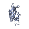
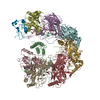
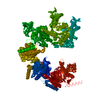
| Title | Cryo-EM structure of Type I-F CRISPR crRNA-guided Csy surveillance complex with bound anti-CRISPR protein AcrF1 | |||||||||||||||||||||||||||
 Components Components |
| |||||||||||||||||||||||||||
 Keywords Keywords | IMMUNE SYSTEM/HYDROLASE/RNA / CRISPR-Cas / IMMUNE SYSTEM-HYDROLASE-RNA complex | |||||||||||||||||||||||||||
| Function / homology |  Function and homology information Function and homology informationmaintenance of CRISPR repeat elements / endonuclease activity / defense response to virus / Hydrolases; Acting on ester bonds / RNA binding Similarity search - Function | |||||||||||||||||||||||||||
| Biological species |   Pseudomonas phage JBD30 (virus) Pseudomonas phage JBD30 (virus) | |||||||||||||||||||||||||||
| Method | ELECTRON MICROSCOPY / single particle reconstruction / cryo EM / Resolution: 3.1 Å | |||||||||||||||||||||||||||
 Authors Authors | Guo, T.W. / Bartesaghi, A. / Yang, H. / Falconieri, V. / Rao, P. / Merk, A. / Fox, T. / Earl, L. / Patel, D.J. / Subramaniam, S. | |||||||||||||||||||||||||||
 Citation Citation |  Journal: Cell / Year: 2017 Journal: Cell / Year: 2017Title: Cryo-EM Structures Reveal Mechanism and Inhibition of DNA Targeting by a CRISPR-Cas Surveillance Complex. Authors: Tai Wei Guo / Alberto Bartesaghi / Hui Yang / Veronica Falconieri / Prashant Rao / Alan Merk / Edward T Eng / Ashleigh M Raczkowski / Tara Fox / Lesley A Earl / Dinshaw J Patel / Sriram Subramaniam /  Abstract: Prokaryotic cells possess CRISPR-mediated adaptive immune systems that protect them from foreign genetic elements, such as invading viruses. A central element of this immune system is an RNA-guided ...Prokaryotic cells possess CRISPR-mediated adaptive immune systems that protect them from foreign genetic elements, such as invading viruses. A central element of this immune system is an RNA-guided surveillance complex capable of targeting non-self DNA or RNA for degradation in a sequence- and site-specific manner analogous to RNA interference. Although the complexes display considerable diversity in their composition and architecture, many basic mechanisms underlying target recognition and cleavage are highly conserved. Using cryoelectron microscopy (cryo-EM), we show that the binding of target double-stranded DNA (dsDNA) to a type I-F CRISPR system yersinia (Csy) surveillance complex leads to large quaternary and tertiary structural changes in the complex that are likely necessary in the pathway leading to target dsDNA degradation by a trans-acting helicase-nuclease. Comparison of the structure of the surveillance complex before and after dsDNA binding, or in complex with three virally encoded anti-CRISPR suppressors that inhibit dsDNA binding, reveals mechanistic details underlying target recognition and inhibition. | |||||||||||||||||||||||||||
| History |
|
- Structure visualization
| Movie |
 Movie viewer Movie viewer |
|---|---|
| Structure viewer | Molecule:  Molmil Molmil Jmol/JSmol Jmol/JSmol |
- Downloads & links
- Download
| PDBx/mmCIF format |  6b46.cif.gz 6b46.cif.gz | 427.2 KB | Display |  PDBx/mmCIF format PDBx/mmCIF format |
|---|---|---|---|---|
| PDB format |  pdb6b46.ent.gz pdb6b46.ent.gz | 343.5 KB | Display |  PDB format PDB format |
| PDBx/mmJSON format |  6b46.json.gz 6b46.json.gz | Tree view |  PDBx/mmJSON format PDBx/mmJSON format | |
| Others |  Other downloads Other downloads |
-Validation report
| Summary document |  6b46_validation.pdf.gz 6b46_validation.pdf.gz | 1 MB | Display |  wwPDB validaton report wwPDB validaton report |
|---|---|---|---|---|
| Full document |  6b46_full_validation.pdf.gz 6b46_full_validation.pdf.gz | 1.1 MB | Display | |
| Data in XML |  6b46_validation.xml.gz 6b46_validation.xml.gz | 60.5 KB | Display | |
| Data in CIF |  6b46_validation.cif.gz 6b46_validation.cif.gz | 96.1 KB | Display | |
| Arichive directory |  https://data.pdbj.org/pub/pdb/validation_reports/b4/6b46 https://data.pdbj.org/pub/pdb/validation_reports/b4/6b46 ftp://data.pdbj.org/pub/pdb/validation_reports/b4/6b46 ftp://data.pdbj.org/pub/pdb/validation_reports/b4/6b46 | HTTPS FTP |
-Related structure data
| Related structure data |  7050MC  7048C  7049C  7051C  7052C  6anvC  6anwC  6b44C  6b45C  6b47C  6b48C M: map data used to model this data C: citing same article ( |
|---|---|
| Similar structure data |
- Links
Links
- Assembly
| Deposited unit | 
|
|---|---|
| 1 |
|
- Components
| #1: Protein | Mass: 37781.547 Da / Num. of mol.: 6 Source method: isolated from a genetically manipulated source Source: (gene. exp.)  Pseudomonas aeruginosa (strain UCBPP-PA14) (bacteria) Pseudomonas aeruginosa (strain UCBPP-PA14) (bacteria)Strain: UCBPP-PA14 / Gene: csy3, csy1-3, PA14_33310 / Plasmid: pCDF-Duet / Production host:  #2: Protein | Mass: 8969.060 Da / Num. of mol.: 2 Source method: isolated from a genetically manipulated source Source: (gene. exp.)  Pseudomonas phage JBD30 (virus) / Gene: JBD30_035 / Plasmid: pRSF-Duet-SUMO / Production host: Pseudomonas phage JBD30 (virus) / Gene: JBD30_035 / Plasmid: pRSF-Duet-SUMO / Production host:  #3: Protein | | Mass: 21629.777 Da / Num. of mol.: 1 Source method: isolated from a genetically manipulated source Source: (gene. exp.)  Pseudomonas aeruginosa (strain UCBPP-PA14) (bacteria) Pseudomonas aeruginosa (strain UCBPP-PA14) (bacteria)Strain: UCBPP-PA14 / Gene: cas6f, csy4, PA14_33300 / Plasmid: pACY-Duet / Production host:  References: UniProt: Q02MM2, Hydrolases; Acting on ester bonds #4: RNA chain | | Mass: 19265.404 Da / Num. of mol.: 1 Source method: isolated from a genetically manipulated source Source: (gene. exp.)   Has protein modification | N | |
|---|
-Experimental details
-Experiment
| Experiment | Method: ELECTRON MICROSCOPY |
|---|---|
| EM experiment | Aggregation state: PARTICLE / 3D reconstruction method: single particle reconstruction |
- Sample preparation
| Component | Name: Type I-F CRISPR crRNA-guided Csy surveillance complex with bound anti-CRISPR protein AcrF1 Type: COMPLEX / Entity ID: all / Source: RECOMBINANT |
|---|---|
| Molecular weight | Value: 0.350 MDa / Experimental value: NO |
| Source (natural) | Organism:  |
| Source (recombinant) | Organism:  |
| Buffer solution | pH: 7.2 Details: 10 mM HEPES, pH 7.2, 150 mM NaCl, 2nM MgCl2, 1 mM DTT |
| Specimen | Conc.: 1 mg/ml / Embedding applied: NO / Shadowing applied: NO / Staining applied: NO / Vitrification applied: YES |
| Specimen support | Grid material: COPPER / Grid mesh size: 200 divisions/in. / Grid type: Quantifoil R1.2/1.3 |
| Vitrification | Instrument: FEI VITROBOT MARK IV / Cryogen name: ETHANE / Humidity: 98 % |
- Electron microscopy imaging
| Experimental equipment |  Model: Titan Krios / Image courtesy: FEI Company |
|---|---|
| Microscopy | Model: FEI TITAN KRIOS |
| Electron gun | Electron source:  FIELD EMISSION GUN / Accelerating voltage: 300 kV / Illumination mode: FLOOD BEAM FIELD EMISSION GUN / Accelerating voltage: 300 kV / Illumination mode: FLOOD BEAM |
| Electron lens | Mode: BRIGHT FIELD |
| Specimen holder | Cryogen: NITROGEN / Specimen holder model: FEI TITAN KRIOS AUTOGRID HOLDER |
| Image recording | Average exposure time: 15.2 sec. / Electron dose: 40 e/Å2 / Detector mode: SUPER-RESOLUTION / Film or detector model: GATAN K2 QUANTUM (4k x 4k) / Num. of real images: 1975 |
| Image scans | Movie frames/image: 38 |
- Processing
| Software | Name: PHENIX / Version: 1.12_2829: / Classification: refinement | ||||||||||||||||||||||||||||
|---|---|---|---|---|---|---|---|---|---|---|---|---|---|---|---|---|---|---|---|---|---|---|---|---|---|---|---|---|---|
| EM software |
| ||||||||||||||||||||||||||||
| CTF correction | Type: PHASE FLIPPING AND AMPLITUDE CORRECTION | ||||||||||||||||||||||||||||
| Symmetry | Point symmetry: C1 (asymmetric) | ||||||||||||||||||||||||||||
| 3D reconstruction | Resolution: 3.1 Å / Resolution method: FSC 0.143 CUT-OFF / Num. of particles: 55966 / Symmetry type: POINT | ||||||||||||||||||||||||||||
| Refine LS restraints |
|
 Movie
Movie Controller


 PDBj
PDBj


