National Institutes of Health/National Institute on Drug Abuse (NIH/NIDA)
GM104962
United States
Memorial Sloan-Kettering Cancer Center Core Grant
P30CA008748
United States
Citation
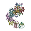
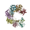
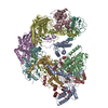
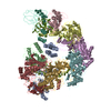
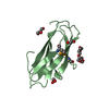
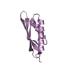
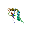
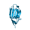
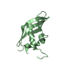
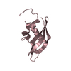
Journal: Cell / Year: 2017 Title: Cryo-EM Structures Reveal Mechanism and Inhibition of DNA Targeting by a CRISPR-Cas Surveillance Complex. Authors: Tai Wei Guo / Alberto Bartesaghi / Hui Yang / Veronica Falconieri / Prashant Rao / Alan Merk / Edward T Eng / Ashleigh M Raczkowski / Tara Fox / Lesley A Earl / Dinshaw J Patel / Sriram Subramaniam / Abstract: Prokaryotic cells possess CRISPR-mediated adaptive immune systems that protect them from foreign genetic elements, such as invading viruses. A central element of this immune system is an RNA-guided ...Prokaryotic cells possess CRISPR-mediated adaptive immune systems that protect them from foreign genetic elements, such as invading viruses. A central element of this immune system is an RNA-guided surveillance complex capable of targeting non-self DNA or RNA for degradation in a sequence- and site-specific manner analogous to RNA interference. Although the complexes display considerable diversity in their composition and architecture, many basic mechanisms underlying target recognition and cleavage are highly conserved. Using cryoelectron microscopy (cryo-EM), we show that the binding of target double-stranded DNA (dsDNA) to a type I-F CRISPR system yersinia (Csy) surveillance complex leads to large quaternary and tertiary structural changes in the complex that are likely necessary in the pathway leading to target dsDNA degradation by a trans-acting helicase-nuclease. Comparison of the structure of the surveillance complex before and after dsDNA binding, or in complex with three virally encoded anti-CRISPR suppressors that inhibit dsDNA binding, reveals mechanistic details underlying target recognition and inhibition.
In the structure databanks used in Yorodumi, some data are registered as the other names, "COVID-19 virus" and "2019-nCoV". Here are the details of the virus and the list of structure data.
Jan 31, 2019. EMDB accession codes are about to change! (news from PDBe EMDB page)
EMDB accession codes are about to change! (news from PDBe EMDB page)
The allocation of 4 digits for EMDB accession codes will soon come to an end. Whilst these codes will remain in use, new EMDB accession codes will include an additional digit and will expand incrementally as the available range of codes is exhausted. The current 4-digit format prefixed with “EMD-” (i.e. EMD-XXXX) will advance to a 5-digit format (i.e. EMD-XXXXX), and so on. It is currently estimated that the 4-digit codes will be depleted around Spring 2019, at which point the 5-digit format will come into force.
The EM Navigator/Yorodumi systems omit the EMD- prefix.
Related info.:Q: What is EMD? / ID/Accession-code notation in Yorodumi/EM Navigator
Yorodumi is a browser for structure data from EMDB, PDB, SASBDB, etc.
This page is also the successor to EM Navigator detail page, and also detail information page/front-end page for Omokage search.
The word "yorodu" (or yorozu) is an old Japanese word meaning "ten thousand". "mi" (miru) is to see.
Related info.:EMDB / PDB / SASBDB / Comparison of 3 databanks / Yorodumi Search / Aug 31, 2016. New EM Navigator & Yorodumi / Yorodumi Papers / Jmol/JSmol / Function and homology information / Changes in new EM Navigator and Yorodumi
 Movie
Movie Controller


 Open data
 Basic information
 Components
 Keywords
 Function and homology information
 Shewanella xiamenensis (bacteria)
 X-RAY DIFFRACTION /
  Authors
 United States, 2items
  Citation
 Structure visualization
 Downloads & links
 Other downloads
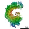


 PDBj
PDBj Assembly
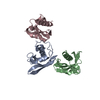

 Mass: 18.015 Da / Num. of mol.: 13 / Source method: isolated from a natural source / Formula: H2O
 Sample preparation
 Processing