 Movie
Movie Controller


+ Open data

- Basic information
| Entry | Database: PDB / ID: 4uij | |||||||||
|---|---|---|---|---|---|---|---|---|---|---|
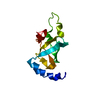
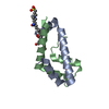
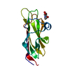
| Title | Crystal structure of the BTB domain of KCTD13 | |||||||||
 Components Components | BTB/POZ DOMAIN-CONTAINING ADAPTER FOR CUL3-MEDIATED RHOA DEGRADATION PROTEIN 1 | |||||||||
 Keywords Keywords | SIGNALING PROTEIN | |||||||||
| Function / homology |  Function and homology information Function and homology informationRND2 GTPase cycle / negative regulation of Rho protein signal transduction / RND3 GTPase cycle / stress fiber assembly / Cul3-RING ubiquitin ligase complex / positive regulation of synaptic transmission / positive regulation of DNA replication / protein homooligomerization / small GTPase binding / cell migration ...RND2 GTPase cycle / negative regulation of Rho protein signal transduction / RND3 GTPase cycle / stress fiber assembly / Cul3-RING ubiquitin ligase complex / positive regulation of synaptic transmission / positive regulation of DNA replication / protein homooligomerization / small GTPase binding / cell migration / proteasome-mediated ubiquitin-dependent protein catabolic process / nuclear body / protein ubiquitination / protein domain specific binding / nucleoplasm / identical protein binding / cytosol Similarity search - Function | |||||||||
| Biological species |  HOMO SAPIENS (human) HOMO SAPIENS (human) | |||||||||
| Method |  X-RAY DIFFRACTION / SYNCHROTRON / MOLECULAR REPLACEMENT / Resolution: 2.7 Å X-RAY DIFFRACTION / SYNCHROTRON / MOLECULAR REPLACEMENT / Resolution: 2.7 Å | |||||||||
 Authors Authors | Pinkas, D.M. / Sanvitale, C.E. / Sorell, F.J. / Solcan, N. / Goubin, S. / Canning, P. / Williams, E. / Chaikuad, A. / Dixon Clarke, S.E. / Tallant, C. ...Pinkas, D.M. / Sanvitale, C.E. / Sorell, F.J. / Solcan, N. / Goubin, S. / Canning, P. / Williams, E. / Chaikuad, A. / Dixon Clarke, S.E. / Tallant, C. / Fonseca, M. / Chalk, R. / Doutch, J. / Krojer, T. / Burgess-Brown, N.A. / von Delft, F. / Arrowsmith, C.H. / Edwards, A.M. / Bountra, C. / Bullock, A. | |||||||||
 Citation Citation | Journal: Biochem. J. / Year: 2017 Title: Structural complexity in the KCTD family of Cullin3-dependent E3 ubiquitin ligases. Authors: Pinkas, D.M. / Sanvitale, C.E. / Bufton, J.C. / Sorrell, F.J. / Solcan, N. / Chalk, R. / Doutch, J. / Bullock, A.N. | |||||||||
| History |
|
- Structure visualization
| Structure viewer | Molecule: MolmilJmol/JSmol |
|---|
- Downloads & links
-Download
| PDBx/mmCIF format | 4uij.cif.gz | 159.3 KB | Display | PDBx/mmCIF format |
|---|---|---|---|---|
| PDB format | pdb4uij.ent.gz | 126.8 KB | Display | PDB format |
| PDBx/mmJSON format | 4uij.json.gz | Tree view | PDBx/mmJSON format | |
| Others |  Other downloads Other downloads |
-Validation report
| Arichive directory | https://data.pdbj.org/pub/pdb/validation_reports/ui/4uijftp://data.pdbj.org/pub/pdb/validation_reports/ui/4uij | HTTPS FTP |
|---|
-Related structure data
| Related structure data | 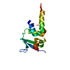 4crhC 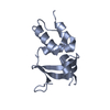 5a15C 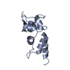 5a6rC 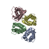 5ftaC  4ues S: Starting model for refinement C: citing same article ( |
|---|---|
| Similar structure data |


-Links
 PDBj
PDBj


- Assembly
| Deposited unit | 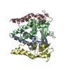
| ||||||||
|---|---|---|---|---|---|---|---|---|---|
| 1 | 
| ||||||||
| 2 | 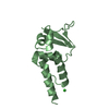
| ||||||||
| 3 | 
| ||||||||
| 4 | 
| ||||||||
| Unit cell |
|
-Components
| #1: Protein | Mass: 15715.845 Da / Num. of mol.: 4 / Fragment: BTB DOMAIN, UNP RESIDUES 27-144 Source method: isolated from a genetically manipulated source Source: (gene. exp.) HOMO SAPIENS (human) / Production host:  #2: Chemical | ChemComp-CL /   Mass: 35.453 Da / Num. of mol.: 4 / Source method: obtained synthetically / Formula: Cl Mass: 35.453 Da / Num. of mol.: 4 / Source method: obtained synthetically / Formula: Cl#3: Water | ChemComp-HOH / |  Mass: 18.015 Da / Num. of mol.: 34 / Source method: isolated from a natural source / Formula: H2O Mass: 18.015 Da / Num. of mol.: 34 / Source method: isolated from a natural source / Formula: H2O |
|---|
-Experimental details
-Experiment
| Experiment | Method: X-RAY DIFFRACTION / Number of used crystals: 1 |
|---|
- Sample preparation
| Crystal | Density Matthews: 1.94 Å3/Da / Density % sol: 36.74 % / Description: NONE |
|---|---|
| Crystal grow | pH: 6.5 / Details: 1.6M MAGNESIUM SULFATE, 0.1M MES PH 6.5 |
-Data collection
| Diffraction | Mean temperature: 100 K |
|---|---|
| Diffraction source | Source: SYNCHROTRON / Site: Diamond  / Beamline: I03 / Wavelength: 0.97625 / Beamline: I03 / Wavelength: 0.97625 |
| Detector | Type: DECTRIS PIXEL / Detector: PIXEL / Date: May 23, 2014 |
| Radiation | Protocol: SINGLE WAVELENGTH / Monochromatic (M) / Laue (L): M / Scattering type: x-ray |
| Radiation wavelength | Wavelength: 0.97625 Å / Relative weight: 1 |
| Reflection | Resolution: 2.7→37.42 Å / Num. obs: 13431 / % possible obs: 99.8 % / Observed criterion σ(I): -1 / Redundancy: 5 % / Biso Wilson estimate: 57.35 Å2 / Rmerge(I) obs: 0.07 / Net I/σ(I): 8.3 |
| Reflection shell | Resolution: 2.7→2.83 Å / Redundancy: 5 % / Mean I/σ(I) obs: 2 / % possible all: 99.8 |
- Processing
| Software |
| ||||||||||||||||||||||||||||||||||||||||||
|---|---|---|---|---|---|---|---|---|---|---|---|---|---|---|---|---|---|---|---|---|---|---|---|---|---|---|---|---|---|---|---|---|---|---|---|---|---|---|---|---|---|---|---|
| Refinement | Method to determine structure: MOLECULAR REPLACEMENT Starting model: PDB ENTRY 4UES 4ues Resolution: 2.7→37.417 Å / SU ML: 0.39 / σ(F): 1.35 / Phase error: 29.22 / Stereochemistry target values: ML
| ||||||||||||||||||||||||||||||||||||||||||
| Solvent computation | Shrinkage radii: 0.9 Å / VDW probe radii: 1.11 Å / Solvent model: FLAT BULK SOLVENT MODEL | ||||||||||||||||||||||||||||||||||||||||||
| Refinement step | Cycle: LAST / Resolution: 2.7→37.417 Å
| ||||||||||||||||||||||||||||||||||||||||||
| Refine LS restraints |
| ||||||||||||||||||||||||||||||||||||||||||
| LS refinement shell |
|
