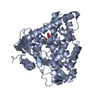
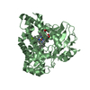
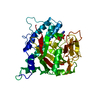
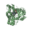
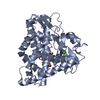
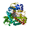
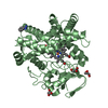
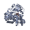
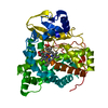
OXIDOREDUCTASE / Cytochrome P450 Oxidase / Haem Protein
Function / homology
Function and homology information
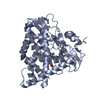
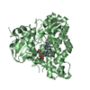
pulcherriminic acid synthase / oxidoreductase activity, acting on paired donors, with incorporation or reduction of molecular oxygen, reduced iron-sulfur protein as one donor, and incorporation of one atom of oxygen / pigment biosynthetic process / monooxygenase activity / iron ion binding / heme binding Similarity search - Function
Type: MARMOSAIC 225 mm CCD / Detector: CCD / Date: Dec 11, 2009
Radiation
Monochromator: SI(111) / Protocol: SINGLE WAVELENGTH / Monochromatic (M) / Laue (L): M / Scattering type: x-ray
Radiation wavelength
Wavelength: 0.979 Å / Relative weight: 1
Reflection
Resolution: 3.1→48.06 Å / Num. all: 18706 / Num. obs: 18488 / % possible obs: 94.3 % / Observed criterion σ(I): 3 / Redundancy: 4.8 % / Biso Wilson estimate: 72.8 Å2 / Rmerge(I) obs: 0.11 / Net I/σ(I): 12.9
Reflection shell
Resolution: 3.1→3.18 Å / Redundancy: 4.9 % / Rmerge(I) obs: 0.43 / Mean I/σ(I) obs: 4 / Num. unique all: 1340 / % possible all: 86.1
-
Processing
Software
Name
Version
Classification
XDS
datascaling
SHARP
phasing
REFMAC
5.5.0070
refinement
XDS
datareduction
XSCALE
datascaling
Refinement
Method to determine structure: SAD / Resolution: 3.1→43.84 Å / Cor.coef. Fo:Fc: 0.929 / Cor.coef. Fo:Fc free: 0.879 / SU B: 45.503 / SU ML: 0.419 / Cross valid method: THROUGHOUT / ESU R Free: 0.519 / Stereochemistry target values: MAXIMUM LIKELIHOOD / Details: HYDROGENS HAVE BEEN ADDED IN THE RIDING POSITIONS
Rfactor
Num. reflection
% reflection
Selection details
Rfree
0.27839
912
4.9 %
RANDOM
Rwork
0.21412
-
-
-
obs
0.21733
18488
100 %
-
all
-
18706
-
-
Solvent computation
Ion probe radii: 0.8 Å / Shrinkage radii: 0.8 Å / VDW probe radii: 1.4 Å / Solvent model: MASK
Displacement parameters
Biso mean: 62.566 Å2
Baniso -1
Baniso -2
Baniso -3
1-
0 Å2
0 Å2
0 Å2
2-
-
0 Å2
0 Å2
3-
-
-
0 Å2
Refinement step
Cycle: LAST / Resolution: 3.1→43.84 Å
Protein
Nucleic acid
Ligand
Solvent
Total
Num. atoms
6029
0
109
5
6143
Refine LS restraints
Refine-ID
Type
Dev ideal
Dev ideal target
Number
X-RAY DIFFRACTION
r_bond_refined_d
0.011
0.022
6281
X-RAY DIFFRACTION
r_bond_other_d
X-RAY DIFFRACTION
r_angle_refined_deg
1.49
2.011
8537
X-RAY DIFFRACTION
r_angle_other_deg
X-RAY DIFFRACTION
r_dihedral_angle_1_deg
6.485
5
760
X-RAY DIFFRACTION
r_dihedral_angle_2_deg
30.439
24.014
294
X-RAY DIFFRACTION
r_dihedral_angle_3_deg
18.571
15
1064
X-RAY DIFFRACTION
r_dihedral_angle_4_deg
20.406
15
45
X-RAY DIFFRACTION
r_chiral_restr
0.099
0.2
944
X-RAY DIFFRACTION
r_gen_planes_refined
0.006
0.021
4783
X-RAY DIFFRACTION
r_gen_planes_other
X-RAY DIFFRACTION
r_nbd_refined
X-RAY DIFFRACTION
r_nbd_other
X-RAY DIFFRACTION
r_nbtor_refined
X-RAY DIFFRACTION
r_nbtor_other
X-RAY DIFFRACTION
r_xyhbond_nbd_refined
X-RAY DIFFRACTION
r_xyhbond_nbd_other
X-RAY DIFFRACTION
r_metal_ion_refined
X-RAY DIFFRACTION
r_metal_ion_other
X-RAY DIFFRACTION
r_symmetry_vdw_refined
X-RAY DIFFRACTION
r_symmetry_vdw_other
X-RAY DIFFRACTION
r_symmetry_hbond_refined
X-RAY DIFFRACTION
r_symmetry_hbond_other
X-RAY DIFFRACTION
r_symmetry_metal_ion_refined
X-RAY DIFFRACTION
r_symmetry_metal_ion_other
X-RAY DIFFRACTION
r_mcbond_it
0.3
1.5
3814
X-RAY DIFFRACTION
r_mcbond_other
X-RAY DIFFRACTION
r_mcangle_it
0.586
2
6164
X-RAY DIFFRACTION
r_scbond_it
1.069
3
2467
X-RAY DIFFRACTION
r_scangle_it
1.873
4.5
2372
X-RAY DIFFRACTION
r_rigid_bond_restr
X-RAY DIFFRACTION
r_sphericity_free
X-RAY DIFFRACTION
r_sphericity_bonded
Refine LS restraints NCS
Auth asym-ID: A / Refine-ID: X-RAY DIFFRACTION
Ens-ID
Dom-ID
Number
Type
Rms dev position (Å)
Weight position
1
1
2946
mediumpositional
0.52
0.5
2
2
43
mediumpositional
0.27
0.5
1
1
2946
mediumthermal
0.49
2
2
2
43
mediumthermal
1.12
2
LS refinement shell
Resolution: 3.1→3.18 Å / Total num. of bins used: 20
Rfactor
Num. reflection
% reflection
Rfree
0.323
64
-
Rwork
0.229
1260
-
obs
-
1260
100 %
Refinement TLS params.
Method: refined / Refine-ID: X-RAY DIFFRACTION
ID
L11 (°2)
L12 (°2)
L13 (°2)
L22 (°2)
L23 (°2)
L33 (°2)
S11 (Å °)
S12 (Å °)
S13 (Å °)
S21 (Å °)
S22 (Å °)
S23 (Å °)
S31 (Å °)
S32 (Å °)
S33 (Å °)
T11 (Å2)
T12 (Å2)
T13 (Å2)
T22 (Å2)
T23 (Å2)
T33 (Å2)
Origin x (Å)
Origin y (Å)
Origin z (Å)
1
6.1361
1.1831
0.7804
4.196
0.0755
3.423
-0.0319
0.0356
-0.4811
0.2795
-0.2797
0.5078
0.717
-0.7524
0.3115
0.6375
-0.1658
0.2725
0.9696
-0.0808
0.2337
18.9921
-15.9982
-8.8502
2
3.567
-0.0694
2.7483
9.6384
3.0048
3.45
0.1856
0.2266
-0.2428
-0.2766
0.2543
-0.4705
-0.2028
0.1502
-0.4398
0.2373
0.0682
0.0773
0.5994
0.0144
0.0691
46.4533
-11.0145
-20.1869
3
2.0054
-0.1221
-0.0282
1.6486
1.4418
3.61
0.1096
0.1293
-0.5582
0.1513
-0.1104
0.2149
0.5629
-0.2276
0.0008
0.4028
-0.0117
0.0166
0.618
-0.0245
0.1989
33.4093
-17.3314
-17.9062
4
6.2246
1.0034
0.7701
7.2836
3.5931
5.0004
0.0525
-0.5829
-0.5673
1.1711
-0.3794
1.1498
1.243
-0.8394
0.3269
0.7586
-0.2793
0.2364
0.8269
0.0001
0.2977
23.0773
-13.6452
-0.6439
5
9.0022
1.1705
1.6643
5.4909
0.2163
7.0963
0.1766
-0.0512
0.5687
0.3438
-0.2302
-0.2129
-0.3728
-0.1277
0.0536
0.2575
0.0218
0.0608
0.5516
-0.0165
0.0733
39.1435
-3.2194
-12.5642
6
7.1593
0.038
-0.9605
1.4971
2.3825
4.813
0.1512
-0.3019
0.3508
0.0323
0.1691
-0.5035
-0.1305
0.6313
-0.3202
0.4251
-0.1014
-0.0077
0.8431
0.0681
0.5088
50.6451
11.6638
18.5635
7
0.1069
-0.0465
-0.1242
8.5388
1.2448
4.8669
-0.0615
-0.0637
-0.2488
-1.1103
0.3698
0.1478
0.5978
0.03
-0.3082
0.6697
-0.073
0.0472
0.761
0.0404
0.68
27.7255
-4.4354
21.7263
8
4.669
0.2522
0.8943
3.685
-1.1702
3.7962
-0.0129
-0.4094
0.1152
0.4604
0.3678
0.4523
-0.4506
-0.2709
-0.3548
0.3071
0.0358
0.0722
0.6107
0.0279
0.0736
23.1153
12.5545
32.2909
9
2.9024
0.1918
-0.5767
1.42
-0.4045
3.4355
0.0874
0.0063
-0.4084
-0.0393
0.2026
0.1221
0.1638
-0.1468
-0.29
0.3236
0.0049
-0.027
0.5759
0.0375
0.1074
31.1176
6.5107
26.4935
10
6.363
-0.7311
-0.2596
4.0797
0.1023
3.5795
0.1739
-0.0178
0.3562
-0.5787
0.0935
-0.0249
-0.2876
0.3171
-0.2674
0.3584
-0.0245
0.0617
0.5613
0.0042
0.0436
34.6344
15.164
16.0611
Refinement TLS group
ID
Refine-ID
Refine TLS-ID
Auth asym-ID
Auth seq-ID
1
X-RAY DIFFRACTION
1
A
5 - 102
2
X-RAY DIFFRACTION
2
A
103 - 150
3
X-RAY DIFFRACTION
3
A
151 - 290
4
X-RAY DIFFRACTION
4
A
291 - 353
5
X-RAY DIFFRACTION
5
A
354 - 403
6
X-RAY DIFFRACTION
6
B
4 - 66
7
X-RAY DIFFRACTION
7
B
67 - 104
8
X-RAY DIFFRACTION
8
B
105 - 168
9
X-RAY DIFFRACTION
9
B
169 - 290
10
X-RAY DIFFRACTION
10
B
291 - 404
+
About Yorodumi
-
News
-
Feb 9, 2022. New format data for meta-information of EMDB entries
New format data for meta-information of EMDB entries
Version 3 of the EMDB header file is now the official format.
The previous official version 1.9 will be removed from the archive.
In the structure databanks used in Yorodumi, some data are registered as the other names, "COVID-19 virus" and "2019-nCoV". Here are the details of the virus and the list of structure data.
Jan 31, 2019. EMDB accession codes are about to change! (news from PDBe EMDB page)
EMDB accession codes are about to change! (news from PDBe EMDB page)
The allocation of 4 digits for EMDB accession codes will soon come to an end. Whilst these codes will remain in use, new EMDB accession codes will include an additional digit and will expand incrementally as the available range of codes is exhausted. The current 4-digit format prefixed with “EMD-” (i.e. EMD-XXXX) will advance to a 5-digit format (i.e. EMD-XXXXX), and so on. It is currently estimated that the 4-digit codes will be depleted around Spring 2019, at which point the 5-digit format will come into force.
The EM Navigator/Yorodumi systems omit the EMD- prefix.
Related info.:Q: What is EMD? / ID/Accession-code notation in Yorodumi/EM Navigator
Yorodumi is a browser for structure data from EMDB, PDB, SASBDB, etc.
This page is also the successor to EM Navigator detail page, and also detail information page/front-end page for Omokage search.
The word "yorodu" (or yorozu) is an old Japanese word meaning "ten thousand". "mi" (miru) is to see.
Related info.:EMDB / PDB / SASBDB / Comparison of 3 databanks / Yorodumi Search / Aug 31, 2016. New EM Navigator & Yorodumi / Yorodumi Papers / Jmol/JSmol / Function and homology information / Changes in new EM Navigator and Yorodumi
 Movie
Movie Controller


 Open data
 Basic information
 Components
 Keywords
 Function and homology information
 X-RAY DIFFRACTION /
  Authors
 Citation
 Structure visualization
 Downloads & links
 Other downloads

 PDBj
PDBj
 Assembly
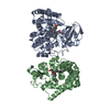

 Mass: 616.487 Da / Num. of mol.: 2 / Source method: obtained synthetically / Formula: C34H32FeN4O4
 Mass: 144.173 Da / Num. of mol.: 2 / Source method: obtained synthetically / Formula: C9H8N2
 Mass: 24.305 Da / Num. of mol.: 1 / Source method: obtained synthetically / Formula: Mg
 Mass: 18.015 Da / Num. of mol.: 5 / Source method: isolated from a natural source / Formula: H2O
 Sample preparation
 / Beamline: X10SA / Wavelength: 0.979 Å
 Processing