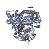
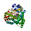
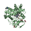
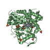
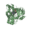
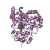
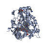
Entry Database : PDB / ID : 5y1iTitle The crystal structure of GfsF Cytochrome P450 Keywords / / Function / homology Function Domain/homology Component
/ / / / / / / / / / / / Biological species Streptomyces graminofaciens (bacteria)Method / / / Resolution : 2 Å Authors Miyanaga, A. / Kudo, F. / Eguchi, T. Funding support Organization Grant number Country Agricultural Chemical Research Foundation 40-5
Journal : Chembiochem / Year : 2017Title : Substrate Recognition by a Dual-Function P450 Monooxygenase GfsF Involved in FD-891 BiosynthesisAuthors : Miyanaga, A. / Takayanagi, R. / Furuya, T. / Kawamata, A. / Itagaki, T. / Iwabuchi, Y. / Kanoh, N. / Kudo, F. / Eguchi, T. History Deposition Jul 20, 2017 Deposition site / Processing site Revision 1.0 Sep 13, 2017 Provider / Type Revision 1.1 Nov 15, 2017 Group / Category Item _citation.journal_volume / _citation.page_first ... _citation.journal_volume / _citation.page_first / _citation.page_last / _citation.title Revision 1.2 Nov 22, 2023 Group Data collection / Database references ... Data collection / Database references / Derived calculations / Refinement description Category chem_comp_atom / chem_comp_bond ... chem_comp_atom / chem_comp_bond / database_2 / pdbx_initial_refinement_model / struct_conn / struct_ncs_dom_lim Item _database_2.pdbx_DOI / _database_2.pdbx_database_accession ... _database_2.pdbx_DOI / _database_2.pdbx_database_accession / _struct_conn.pdbx_dist_value / _struct_conn.ptnr1_auth_asym_id / _struct_conn.ptnr1_auth_comp_id / _struct_conn.ptnr1_auth_seq_id / _struct_conn.ptnr1_label_asym_id / _struct_conn.ptnr1_label_atom_id / _struct_conn.ptnr1_label_comp_id / _struct_conn.ptnr1_label_seq_id / _struct_conn.ptnr2_auth_asym_id / _struct_conn.ptnr2_auth_comp_id / _struct_conn.ptnr2_auth_seq_id / _struct_conn.ptnr2_label_asym_id / _struct_conn.ptnr2_label_atom_id / _struct_conn.ptnr2_label_comp_id / _struct_ncs_dom_lim.beg_auth_comp_id / _struct_ncs_dom_lim.beg_label_asym_id / _struct_ncs_dom_lim.beg_label_comp_id / _struct_ncs_dom_lim.beg_label_seq_id / _struct_ncs_dom_lim.end_auth_comp_id / _struct_ncs_dom_lim.end_label_asym_id / _struct_ncs_dom_lim.end_label_comp_id / _struct_ncs_dom_lim.end_label_seq_id
Show all Show less
 Movie
Movie Controller


 Open data
 Basic information
 Components
 Keywords
 Function and homology information
 Streptomyces graminofaciens (bacteria)
 X-RAY DIFFRACTION /
  Authors
 Japan, 1items
  Citation
 Structure visualization
 Downloads & links
 Other downloads
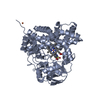
 PDBj
PDBj
 Assembly
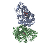
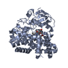
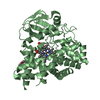

 Mass: 616.487 Da / Num. of mol.: 2 / Source method: obtained synthetically / Formula: C34H32FeN4O4
 Mass: 106.120 Da / Num. of mol.: 2 / Source method: obtained synthetically / Formula: C4H10O3
 Mass: 150.173 Da / Num. of mol.: 1 / Source method: obtained synthetically / Formula: C6H14O4
 Mass: 18.015 Da / Num. of mol.: 392 / Source method: isolated from a natural source / Formula: H2O
 Sample preparation
 Processing