 Movie
Movie Controller

[English] 日本語
 Yorodumi
- PDB-2y46: Structure of the mixed-function P450 MycG in complex with mycinam... -
+ Open data

- Basic information
| Entry | Database: PDB / ID: 2y46 | ||||||
|---|---|---|---|---|---|---|---|
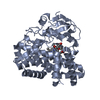
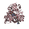
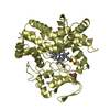
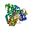
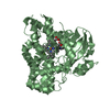
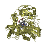
| Title | Structure of the mixed-function P450 MycG in complex with mycinamicin IV in C 2 2 21 space group | ||||||
 Components Components | P-450-LIKE PROTEIN | ||||||
 Keywords Keywords | OXIDOREDUCTASE / MYCINAMICIN BIOSYNTHESIS | ||||||
| Function / homology |  Function and homology information Function and homology informationOxidoreductases; Acting on paired donors, with incorporation or reduction of molecular oxygen / oxidoreductase activity, acting on paired donors, with incorporation or reduction of molecular oxygen / antibiotic biosynthetic process / monooxygenase activity / iron ion binding / heme binding Similarity search - Function | ||||||
| Biological species |  MICROMONOSPORA GRISEORUBIDA (bacteria) MICROMONOSPORA GRISEORUBIDA (bacteria) | ||||||
| Method |  X-RAY DIFFRACTION / SYNCHROTRON / MOLECULAR REPLACEMENT / Resolution: 1.83 Å X-RAY DIFFRACTION / SYNCHROTRON / MOLECULAR REPLACEMENT / Resolution: 1.83 Å | ||||||
 Authors Authors | Li, S. / Kells, P.M. / Sherman, D.H. / Podust, L.M. | ||||||
 Citation Citation | Journal: J.Biol.Chem. / Year: 2012 Title: Substrate Recognition by the Multifunctional Cytochrome P450 Mycg in Mycinamicin Hydroxylation and Epoxidation Reactions. Authors: Li, S. / Tietz, D.R. / Rutaganira, F.U. / Kells, P.M. / Anzai, Y. / Kato, F. / Pochapsky, T.C. / Sherman, D.H. / Podust, L.M. | ||||||
| History |
|
- Structure visualization
| Structure viewer | Molecule: MolmilJmol/JSmol |
|---|
- Downloads & links
-Download
| PDBx/mmCIF format | 2y46.cif.gz | 530 KB | Display | PDBx/mmCIF format |
|---|---|---|---|---|
| PDB format | pdb2y46.ent.gz | 434.6 KB | Display | PDB format |
| PDBx/mmJSON format | 2y46.json.gz | Tree view | PDBx/mmJSON format | |
| Others |  Other downloads Other downloads |
-Validation report
| Arichive directory | https://data.pdbj.org/pub/pdb/validation_reports/y4/2y46ftp://data.pdbj.org/pub/pdb/validation_reports/y4/2y46 | HTTPS FTP |
|---|
-Related structure data
| Related structure data | 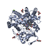 2y5nC 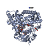 2y5zC 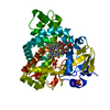 2y98C  2ycaC 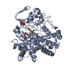 2ygxC  3zsnC  4aw3C  2y4h S: Starting model for refinement C: citing same article ( |
|---|---|
| Similar structure data |


-Links
 PDBj
PDBj

- Assembly
| Deposited unit | 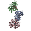
| ||||||||||||||||||||||||||||||||||||
|---|---|---|---|---|---|---|---|---|---|---|---|---|---|---|---|---|---|---|---|---|---|---|---|---|---|---|---|---|---|---|---|---|---|---|---|---|---|
| 1 | 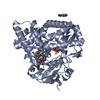
| ||||||||||||||||||||||||||||||||||||
| 2 | 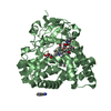
| ||||||||||||||||||||||||||||||||||||
| 3 | 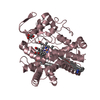
| ||||||||||||||||||||||||||||||||||||
| Unit cell |
| ||||||||||||||||||||||||||||||||||||
| Components on special symmetry positions |
| ||||||||||||||||||||||||||||||||||||
| Noncrystallographic symmetry (NCS) | NCS domain:
NCS domain segments: Component-ID: 1 / Ens-ID: 1 / Beg auth comp-ID: GLU / Beg label comp-ID: GLU / End auth comp-ID: HEM / End label comp-ID: HEM / Refine code: 4 / Auth seq-ID: 5 - 450 / Label seq-ID: 25
NCS oper:
|
-Components
-Protein , 1 types, 3 molecules ABC
| #1: Protein | Mass: 46556.762 Da / Num. of mol.: 3 Source method: isolated from a genetically manipulated source Source: (gene. exp.) MICROMONOSPORA GRISEORUBIDA (bacteria) / Production host: |
|---|
-Non-polymers , 5 types, 931 molecules 


| #2: Chemical |  Mass: 616.487 Da / Num. of mol.: 3 / Source method: obtained synthetically / Formula: C34H32FeN4O4 Mass: 616.487 Da / Num. of mol.: 3 / Source method: obtained synthetically / Formula: C34H32FeN4O4#3: Chemical |  Mass: 695.880 Da / Num. of mol.: 3 / Source method: obtained synthetically / Formula: C37H61NO11 Mass: 695.880 Da / Num. of mol.: 3 / Source method: obtained synthetically / Formula: C37H61NO11#4: Chemical |  Mass: 120.152 Da / Num. of mol.: 3 / Source method: obtained synthetically / Formula: C7H8N2 Mass: 120.152 Da / Num. of mol.: 3 / Source method: obtained synthetically / Formula: C7H8N2#5: Chemical | ChemComp-GOL /  Mass: 92.094 Da / Num. of mol.: 6 / Source method: obtained synthetically / Formula: C3H8O3 Mass: 92.094 Da / Num. of mol.: 6 / Source method: obtained synthetically / Formula: C3H8O3#6: Water | ChemComp-HOH / | Mass: 18.015 Da / Num. of mol.: 916 / Source method: isolated from a natural source / Formula: H2O |
|---|
-Experimental details
-Experiment
| Experiment | Method: X-RAY DIFFRACTION / Number of used crystals: 1 |
|---|
- Sample preparation
| Crystal | Density Matthews: 2.4 Å3/Da / Density % sol: 48.9 % / Description: NONE |
|---|---|
| Crystal grow | Temperature: 296 K / pH: 7 Details: 12% PEG MME 5000, 5% TACSIMATE PH 7.0, 1% BENZAMIDINE HYDROCHLORIDE, TEMP 23 C |
-Data collection
| Diffraction | Mean temperature: 110 K |
|---|---|
| Diffraction source | Source: SYNCHROTRON / Site: ALS  / Beamline: 8.3.1 / Wavelength: 1.11588 / Beamline: 8.3.1 / Wavelength: 1.11588 |
| Detector | Type: MARRESEARCH / Detector: CCD / Date: Jan 4, 2010 / Details: MIRRORS |
| Radiation | Monochromator: SI (111) DOUBLE CRYSTAL / Protocol: SINGLE WAVELENGTH / Monochromatic (M) / Laue (L): M / Scattering type: x-ray |
| Radiation wavelength | Wavelength: 1.11588 Å / Relative weight: 1 |
| Reflection | Resolution: 1.83→147.18 Å / Num. obs: 95693 / % possible obs: 83.1 % / Observed criterion σ(I): 1.5 / Redundancy: 5.3 % / Biso Wilson estimate: 23 Å2 / Rmerge(I) obs: 0.08 / Net I/σ(I): 11.1 |
| Reflection shell | Resolution: 1.83→1.93 Å / Redundancy: 2.4 % / Rmerge(I) obs: 0.56 / Mean I/σ(I) obs: 1.5 / % possible all: 36.2 |
- Processing
| Software |
| ||||||||||||||||||||||||||||||||||||||||||||||||||||||||||||||||||||||||||||||||||||||||||||||||||||||||||||||||||||||||||||||||||||||||||||||||||||||||||||||||||||||||||||||||||||||
|---|---|---|---|---|---|---|---|---|---|---|---|---|---|---|---|---|---|---|---|---|---|---|---|---|---|---|---|---|---|---|---|---|---|---|---|---|---|---|---|---|---|---|---|---|---|---|---|---|---|---|---|---|---|---|---|---|---|---|---|---|---|---|---|---|---|---|---|---|---|---|---|---|---|---|---|---|---|---|---|---|---|---|---|---|---|---|---|---|---|---|---|---|---|---|---|---|---|---|---|---|---|---|---|---|---|---|---|---|---|---|---|---|---|---|---|---|---|---|---|---|---|---|---|---|---|---|---|---|---|---|---|---|---|---|---|---|---|---|---|---|---|---|---|---|---|---|---|---|---|---|---|---|---|---|---|---|---|---|---|---|---|---|---|---|---|---|---|---|---|---|---|---|---|---|---|---|---|---|---|---|---|---|---|
| Refinement | Method to determine structure: MOLECULAR REPLACEMENT Starting model: PDB ENTRY 2Y4H 2y4h Resolution: 1.83→220.77 Å / Cor.coef. Fo:Fc: 0.964 / Cor.coef. Fo:Fc free: 0.928 / SU B: 7.588 / SU ML: 0.101 / Cross valid method: THROUGHOUT / ESU R Free: 0.158 / Stereochemistry target values: MAXIMUM LIKELIHOOD / Details: HYDROGENS HAVE BEEN ADDED IN THE RIDING POSITIONS
| ||||||||||||||||||||||||||||||||||||||||||||||||||||||||||||||||||||||||||||||||||||||||||||||||||||||||||||||||||||||||||||||||||||||||||||||||||||||||||||||||||||||||||||||||||||||
| Solvent computation | Ion probe radii: 0.8 Å / Shrinkage radii: 0.8 Å / VDW probe radii: 1.4 Å / Solvent model: MASK | ||||||||||||||||||||||||||||||||||||||||||||||||||||||||||||||||||||||||||||||||||||||||||||||||||||||||||||||||||||||||||||||||||||||||||||||||||||||||||||||||||||||||||||||||||||||
| Displacement parameters | Biso mean: 23.09 Å2
| ||||||||||||||||||||||||||||||||||||||||||||||||||||||||||||||||||||||||||||||||||||||||||||||||||||||||||||||||||||||||||||||||||||||||||||||||||||||||||||||||||||||||||||||||||||||
| Refinement step | Cycle: LAST / Resolution: 1.83→220.77 Å
| ||||||||||||||||||||||||||||||||||||||||||||||||||||||||||||||||||||||||||||||||||||||||||||||||||||||||||||||||||||||||||||||||||||||||||||||||||||||||||||||||||||||||||||||||||||||
| Refine LS restraints |
|