cholesterol 7alpha-monooxygenase / 24-hydroxycholesterol 7alpha-hydroxylase / cholesterol 7-alpha-monooxygenase activity / 24S-hydroxycholesterol 7-alpha-hydroxylase activity / bile acid biosynthetic process / sterol metabolic process / negative regulation of collagen biosynthetic process / regulation of bile acid biosynthetic process / cellular response to cholesterol / negative regulation of fatty acid biosynthetic process ...cholesterol 7alpha-monooxygenase / 24-hydroxycholesterol 7alpha-hydroxylase / cholesterol 7-alpha-monooxygenase activity / 24S-hydroxycholesterol 7-alpha-hydroxylase activity / bile acid biosynthetic process / sterol metabolic process / negative regulation of collagen biosynthetic process / regulation of bile acid biosynthetic process / cellular response to cholesterol / negative regulation of fatty acid biosynthetic process / cholesterol catabolic process / Synthesis of bile acids and bile salts / intracellular membrane-bounded organelle / Synthesis of bile acids and bile salts via 27-hydroxycholesterol / Endogenous sterols / Synthesis of bile acids and bile salts via 7alpha-hydroxycholesterol / cholesterol homeostasis / cellular response to glucose stimulus / PPARA activates gene expression / positive regulation of cholesterol biosynthetic process / response to ethanol / iron ion binding / heme binding / endoplasmic reticulum membrane Similarity search - Function
Resolution: 1.9→50 Å / Cor.coef. Fo:Fc: 0.963 / Cor.coef. Fo:Fc free: 0.94 / WRfactor Rfree: 0.214 / WRfactor Rwork: 0.173 / Occupancy max: 1 / Occupancy min: 0.3 / SU B: 3.55 / SU ML: 0.098 / Cross valid method: THROUGHOUT / σ(F): 0 / ESU R: 0.134 / ESU R Free: 0.13 / Stereochemistry target values: MAXIMUM LIKELIHOOD Details: HYDROGENS HAVE BEEN ADDED IN THE RIDING POSITIONS U VALUES : REFINED INDIVIDUALLY Geometry restraints for 7-ketocholesterol wer prepared by the PRODRG server. Heme geometry restraints were ...Details: HYDROGENS HAVE BEEN ADDED IN THE RIDING POSITIONS U VALUES : REFINED INDIVIDUALLY Geometry restraints for 7-ketocholesterol wer prepared by the PRODRG server. Heme geometry restraints were modified from the CCP4 default using data from the Cambridge Structural Database, using the Mogul query tool. PHENIX, COOT and the MOLPROBITY server were also used during refinement.
Rfactor
Num. reflection
% reflection
Selection details
Rfree
0.225
2066
2.241 %
THIN SHELLS (SFTOOLS)
Rwork
0.1852
-
-
-
obs
0.186
92179
97.601 %
-
Solvent computation
Ion probe radii: 0.8 Å / Shrinkage radii: 0.8 Å / VDW probe radii: 1.2 Å / Solvent model: MASK BULK SOLVENT
In the structure databanks used in Yorodumi, some data are registered as the other names, "COVID-19 virus" and "2019-nCoV". Here are the details of the virus and the list of structure data.
Jan 31, 2019. EMDB accession codes are about to change! (news from PDBe EMDB page)
EMDB accession codes are about to change! (news from PDBe EMDB page)
The allocation of 4 digits for EMDB accession codes will soon come to an end. Whilst these codes will remain in use, new EMDB accession codes will include an additional digit and will expand incrementally as the available range of codes is exhausted. The current 4-digit format prefixed with “EMD-” (i.e. EMD-XXXX) will advance to a 5-digit format (i.e. EMD-XXXXX), and so on. It is currently estimated that the 4-digit codes will be depleted around Spring 2019, at which point the 5-digit format will come into force.
The EM Navigator/Yorodumi systems omit the EMD- prefix.
Related info.:Q: What is EMD? / ID/Accession-code notation in Yorodumi/EM Navigator
Yorodumi is a browser for structure data from EMDB, PDB, SASBDB, etc.
This page is also the successor to EM Navigator detail page, and also detail information page/front-end page for Omokage search.
The word "yorodu" (or yorozu) is an old Japanese word meaning "ten thousand". "mi" (miru) is to see.
Related info.:EMDB / PDB / SASBDB / Comparison of 3 databanks / Yorodumi Search / Aug 31, 2016. New EM Navigator & Yorodumi / Yorodumi Papers / Jmol/JSmol / Function and homology information / Changes in new EM Navigator and Yorodumi
 Movie
Movie Controller

 Yorodumi
 Open data
 Basic information
 Components
 Keywords
 Function and homology information
 Homo sapiens (human)
 X-RAY DIFFRACTION /
  Authors
 Citation
 Structure visualization
 Downloads & links
 Other downloads


 PDBj
PDBj


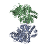
 Assembly
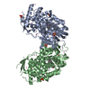


 Mass: 616.487 Da / Num. of mol.: 2 / Source method: obtained synthetically / Formula: C34H32FeN4O4
 Mass: 400.637 Da / Num. of mol.: 2 / Source method: obtained synthetically / Formula: C27H44O2
 Mass: 96.063 Da / Num. of mol.: 6 / Source method: obtained synthetically / Formula: SO4
 Num. of mol.: 14 / Source method: obtained synthetically
 Sample preparation
 / Beamline: 08ID-1 / Wavelength: 0.97625 Å
 Processing