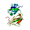
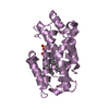
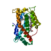
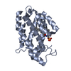
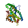
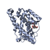
- PDB-5kdi: How FAPP2 Selects Simple Glycosphingolipids Using the GLTP-fold -
+
Open data
ID or keywords:
Loading...
-
Basic information
Entry
Database: PDB / ID: 5kdi
Title
How FAPP2 Selects Simple Glycosphingolipids Using the GLTP-fold
Components
Pleckstrin homology domain-containing family A member 8
Keywords
LIPID TRANSPORT / GLTP-fold / lipid transfer
Function / homology
Function and homology information
ER to Golgi ceramide transport / glycolipid transfer activity / ceramide 1-phosphate transfer activity / ceramide transport / ceramide 1-phosphate binding / glycolipid binding / Glycosphingolipid transport / intermembrane lipid transfer / ceramide binding / phosphatidylinositol-4-phosphate binding ...ER to Golgi ceramide transport / glycolipid transfer activity / ceramide 1-phosphate transfer activity / ceramide transport / ceramide 1-phosphate binding / glycolipid binding / Glycosphingolipid transport / intermembrane lipid transfer / ceramide binding / phosphatidylinositol-4-phosphate binding / lipid transport / Synthesis of PIPs at the plasma membrane / trans-Golgi network / protein transport / Golgi membrane / Golgi apparatus / nucleoplasm / cytosol Similarity search - Function
Glycolipid transfer protein domain / Glycolipid transfer protein superfamily / Glycolipid transfer protein (GLTP) / Glycolipid transfer protein, GLTP / Glycolipid transfer protein / PH domain / PH domain profile. / Pleckstrin homology domain. / Pleckstrin homology domain / PH-like domain superfamily ...Glycolipid transfer protein domain / Glycolipid transfer protein superfamily / Glycolipid transfer protein (GLTP) / Glycolipid transfer protein, GLTP / Glycolipid transfer protein / PH domain / PH domain profile. / Pleckstrin homology domain. / Pleckstrin homology domain / PH-like domain superfamily / Orthogonal Bundle / Mainly Alpha Similarity search - Domain/homology
Pleckstrinhomologydomain-containingfamilyAmember8 / PH domain-containing family A member 8 / Phosphatidylinositol-four-phosphate adapter protein 2 / ...PH domain-containing family A member 8 / Phosphatidylinositol-four-phosphate adapter protein 2 / hFAPP2 / Serologically defined breast cancer antigen NY-BR-86 / FAPP2-GLTPH domain
Mass: 23489.021 Da / Num. of mol.: 2 / Mutation: E377A, E378K Source method: isolated from a genetically manipulated source Source: (gene. exp.) Homo sapiens (human) / Gene: PLEKHA8, FAPP2 / Production host: Escherichia coli (E. coli) / References: UniProt: Q96JA3
In the structure databanks used in Yorodumi, some data are registered as the other names, "COVID-19 virus" and "2019-nCoV". Here are the details of the virus and the list of structure data.
Jan 31, 2019. EMDB accession codes are about to change! (news from PDBe EMDB page)
EMDB accession codes are about to change! (news from PDBe EMDB page)
The allocation of 4 digits for EMDB accession codes will soon come to an end. Whilst these codes will remain in use, new EMDB accession codes will include an additional digit and will expand incrementally as the available range of codes is exhausted. The current 4-digit format prefixed with “EMD-” (i.e. EMD-XXXX) will advance to a 5-digit format (i.e. EMD-XXXXX), and so on. It is currently estimated that the 4-digit codes will be depleted around Spring 2019, at which point the 5-digit format will come into force.
The EM Navigator/Yorodumi systems omit the EMD- prefix.
Related info.:Q: What is EMD? / ID/Accession-code notation in Yorodumi/EM Navigator
Yorodumi is a browser for structure data from EMDB, PDB, SASBDB, etc.
This page is also the successor to EM Navigator detail page, and also detail information page/front-end page for Omokage search.
The word "yorodu" (or yorozu) is an old Japanese word meaning "ten thousand". "mi" (miru) is to see.
Related info.:EMDB / PDB / SASBDB / Comparison of 3 databanks / Yorodumi Search / Aug 31, 2016. New EM Navigator & Yorodumi / Yorodumi Papers / Jmol/JSmol / Function and homology information / Changes in new EM Navigator and Yorodumi
 Movie
Movie Controller


 Open data
 Basic information
 Components
 Keywords
 Function and homology information
 Homo sapiens (human)
 X-RAY DIFFRACTION /
  Authors
 United States,
  Spain,
  Russian Federation, 3items
  Citation
 Structure visualization
 Downloads & links
 Other downloads


 PDBj
PDBj
 Assembly


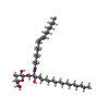
 Mass: 726.079 Da / Num. of mol.: 2 / Source method: obtained synthetically / Formula: C42H79NO8
 Mass: 18.015 Da / Num. of mol.: 493 / Source method: isolated from a natural source / Formula: H2O
 Sample preparation
 / Beamline: ID23-1 / Wavelength: 0.97242 Å
 Processing