[English] 日本語
Yorodumi
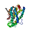
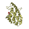
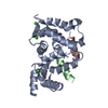
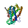
- PDB-1sx6: Crystal structure of human Glycolipid Transfer protein in lactosy... -

+
Open data


ID or keywords:

Loading...

-
Basic information

Entry
Database: PDB / ID: 1sx6
TitleCrystal structure of human Glycolipid Transfer protein in lactosylceramide-bound form
ComponentsGlycolipid transfer protein
KeywordsLIPID TRANSPORT / glycosphingolipid transfer protein-lactosylceramide complex
Function / homology
Function and homology information


Glycosphingolipid biosynthesis / glycolipid transfer activity / lipid transfer activity / ceramide 1-phosphate binding / ceramide 1-phosphate transfer activity / ER to Golgi ceramide transport / glycolipid binding / glycosphingolipid metabolic process / intermembrane lipid transfer / response to immobilization stress ...Glycosphingolipid biosynthesis / glycolipid transfer activity / lipid transfer activity / ceramide 1-phosphate binding / ceramide 1-phosphate transfer activity / ER to Golgi ceramide transport / glycolipid binding / glycosphingolipid metabolic process / intermembrane lipid transfer / response to immobilization stress / lipid binding / identical protein binding / cytosol
Similarity search - Function
Glycolipid transfer protein domain / Glycolipid transfer protein superfamily / Glycolipid transfer protein (GLTP) / Glycolipid transfer protein, GLTP / Glycolipid transfer protein / Orthogonal Bundle / Mainly Alpha
Similarity search - Domain/homology
beta-lactose / N-OCTANE / OLEIC ACID / SPHINGOSINE / Glycolipid transfer protein
Similarity search - Component
Biological speciesHomo sapiens (human)
MethodX-RAY DIFFRACTION / MOLECULAR REPLACEMENT / Resolution: 1.95 Å
AuthorsMalinina, L. / Malakhova, M.L. / Teplov, A. / Brown, R.E. / Patel, D.J.
CitationJournal: Nature / Year: 2004
Title: Structural basis for glycosphingolipid transfer specificity.
Authors: Malinina, L. / Malakhova, M.L. / Teplov, A. / Brown, R.E. / Patel, D.J.
History
DepositionMar 30, 2004Deposition site: RCSB / Processing site: RCSB
Revision 1.0Aug 31, 2004Provider: repository / Type: Initial release
Revision 1.1Apr 30, 2008Group: Version format compliance
Revision 1.2Jul 13, 2011Group: Non-polymer description / Version format compliance
Revision 2.0Jul 29, 2020Group: Advisory / Atomic model ...Advisory / Atomic model / Data collection / Derived calculations / Non-polymer description / Structure summary
Category: atom_site / chem_comp ...atom_site / chem_comp / database_PDB_caveat / entity / entity_name_com / pdbx_branch_scheme / pdbx_chem_comp_identifier / pdbx_entity_branch / pdbx_entity_branch_descriptor / pdbx_entity_branch_link / pdbx_entity_branch_list / pdbx_entity_nonpoly / pdbx_molecule_features / pdbx_nonpoly_scheme / pdbx_validate_chiral / struct_conn / struct_site / struct_site_gen
Item: _atom_site.B_iso_or_equiv / _atom_site.Cartn_x ..._atom_site.B_iso_or_equiv / _atom_site.Cartn_x / _atom_site.Cartn_y / _atom_site.Cartn_z / _atom_site.auth_asym_id / _atom_site.auth_atom_id / _atom_site.auth_comp_id / _atom_site.auth_seq_id / _atom_site.label_atom_id / _atom_site.label_comp_id / _chem_comp.formula / _chem_comp.formula_weight / _chem_comp.id / _chem_comp.mon_nstd_flag / _chem_comp.name / _chem_comp.type / _entity.formula_weight / _entity.pdbx_description / _entity.type / _pdbx_validate_chiral.auth_asym_id / _pdbx_validate_chiral.auth_atom_id / _pdbx_validate_chiral.auth_comp_id / _pdbx_validate_chiral.auth_seq_id
Description: Carbohydrate remediation / Provider: repository / Type: Remediation
Revision 2.1Aug 23, 2023Group: Data collection / Database references ...Data collection / Database references / Derived calculations / Refinement description / Structure summary
Category: chem_comp / chem_comp_atom ...chem_comp / chem_comp_atom / chem_comp_bond / database_2 / pdbx_initial_refinement_model / struct_conn
Item: _chem_comp.pdbx_synonyms / _database_2.pdbx_DOI ..._chem_comp.pdbx_synonyms / _database_2.pdbx_DOI / _database_2.pdbx_database_accession / _struct_conn.pdbx_leaving_atom_flag
Remark 600heterogen The residues LAT, SPH and OLA together form a lactosylceramide. The atom O in SPH and O1 ...heterogen The residues LAT, SPH and OLA together form a lactosylceramide. The atom O in SPH and O1 in OLA are lost during the formation of this ligand.

-
Structure visualization

Structure viewerMolecule:
MolmilJmol/JSmol

Downloads & links

-
Assembly

Deposited unit
A: Glycolipid transfer protein
hetero molecules


Theoretical massNumber of molelcules
Total (without water)24,9165
Polymers23,8781
Non-polymers1,0384
Water2,882160
1


  • Idetical with deposited unit
  • defined by author
TypeNameSymmetry operationNumber
identity operation1_555x,y,z1
Unit cell
Length a, b, c (Å)75.599, 49.073, 68.538
Angle α, β, γ (deg.)90.00, 122.52, 90.00
Int Tables number5
Space group name H-MC121
Components on special symmetry positions
IDModelComponents
11A-319-

HOH

-
Components

-
Protein / Sugars , 2 types, 2 molecules A

#1: Protein Glycolipid transfer protein / / GLTP


Mass: 23877.777 Da / Num. of mol.: 1
Source method: isolated from a genetically manipulated source
Source: (gene. exp.) Homo sapiens (human) / Gene: GLTP / Production host: Escherichia coli (E. coli) / References: UniProt: Q9NZD2
#2: Polysaccharide beta-D-galactopyranose-(1-4)-beta-D-glucopyranose / beta-lactose


Type: oligosaccharide, Oligosaccharide / Class: Nutrient / Mass: 342.297 Da / Num. of mol.: 1
Source method: isolated from a genetically manipulated source
Details: oligosaccharide / References: beta-lactose
DescriptorTypeProgram
DGalpb1-4DGlcpb1-ROHGlycam Condensed SequenceGMML 1.0
WURCS=2.0/2,2,1/[a2122h-1b_1-5][a2112h-1b_1-5]/1-2/a4-b1WURCSPDB2Glycan 1.1.0
[][a-D-Glcp]{[(4+1)][b-D-Galp]{}}LINUCSPDB-CARE

-
Non-polymers , 4 types, 163 molecules

#3: Chemical ChemComp-SPH / SPHINGOSINE / Sphingosine


Mass: 299.492 Da / Num. of mol.: 1 / Source method: obtained synthetically / Formula: C18H37NO2
#4: Chemical ChemComp-OLA / OLEIC ACID / Oleic acid


Mass: 282.461 Da / Num. of mol.: 1 / Source method: obtained synthetically / Formula: C18H34O2
#5: Chemical ChemComp-OCT / N-OCTANE / Octane


Mass: 114.229 Da / Num. of mol.: 1 / Source method: obtained synthetically / Formula: C8H18
#6: Water ChemComp-HOH / water / Water


Mass: 18.015 Da / Num. of mol.: 160 / Source method: isolated from a natural source / Formula: H2O

-
Experimental details

-
Experiment

ExperimentMethod: X-RAY DIFFRACTION / Number of used crystals: 1

-
Sample preparation

CrystalDensity Matthews: 2.24 Å3/Da / Density % sol: 45.18 %
Crystal growTemperature: 298 K / Method: vapor diffusion, hanging drop / pH: 4.5
Details: PEG 8000, potassium phosphate, pH 4.5, VAPOR DIFFUSION, HANGING DROP, temperature 298K

-
Data collection

DiffractionMean temperature: 100 K
Diffraction sourceSource: ROTATING ANODE / Type: RIGAKU RUH3R / Wavelength: 1.5418 Å
DetectorType: RIGAKU RAXIS HTC / Detector: IMAGE PLATE / Date: Dec 19, 2003 / Details: mirrors
RadiationProtocol: SINGLE WAVELENGTH / Monochromatic (M) / Laue (L): M / Scattering type: x-ray
Radiation wavelengthWavelength: 1.5418 Å / Relative weight: 1
ReflectionResolution: 1.9→100 Å / Num. all: 16853 / Num. obs: 16786 / % possible obs: 99.6 % / Observed criterion σ(F): 0 / Observed criterion σ(I): 0 / Redundancy: 4.33 % / Rmerge(I) obs: 0.117 / Net I/σ(I): 15
Reflection shellResolution: 1.9→1.97 Å / Rmerge(I) obs: 0.49 / Mean I/σ(I) obs: 1.7 / Num. unique all: 1672

-
Processing

Software
NameVersionClassification
REFMAC5.1.24refinement
CrystalClear(MSC/RIGAKU)data reduction
SCALEPACKdata scaling
AMoREphasing
RefinementMethod to determine structure: MOLECULAR REPLACEMENT
Starting model: PDB ENTRY 1SWX
Resolution: 1.95→20 Å / Cor.coef. Fo:Fc: 0.966 / Cor.coef. Fo:Fc free: 0.949 / SU B: 6.124 / SU ML: 0.158 / Cross valid method: THROUGHOUT / σ(F): 0 / σ(I): 0 / ESU R: 0.194 / ESU R Free: 0.173 / Stereochemistry target values: MAXIMUM LIKELIHOOD / Details: HYDROGENS HAVE BEEN ADDED IN THE RIDING POSITIONS
RfactorNum. reflection% reflectionSelection details
Rfree0.24429 785 5 %RANDOM
Rwork0.19202 ---
all0.1947 ---
obs0.19479 14761 99.78 %-
Solvent computationIon probe radii: 0.8 Å / Shrinkage radii: 0.8 Å / VDW probe radii: 1.4 Å / Solvent model: BABINET MODEL WITH MASK
Displacement parametersBiso mean: 38.691 Å2
Baniso -1Baniso -2Baniso -3
1--2.33 Å20 Å2-0.23 Å2
2--1.27 Å20 Å2
3---0.81 Å2
Refinement stepCycle: LAST / Resolution: 1.95→20 Å
ProteinNucleic acidLigandSolventTotal
Num. atoms1645 0 70 160 1875
Refine LS restraints
Refine-IDTypeDev idealDev ideal targetNumber
X-RAY DIFFRACTIONr_bond_refined_d0.0110.0221758
X-RAY DIFFRACTIONr_bond_other_d0.0020.021650
X-RAY DIFFRACTIONr_angle_refined_deg1.241.9982365
X-RAY DIFFRACTIONr_angle_other_deg0.84533850
X-RAY DIFFRACTIONr_dihedral_angle_1_deg5.3985203
X-RAY DIFFRACTIONr_chiral_restr0.0710.2267
X-RAY DIFFRACTIONr_gen_planes_refined0.0040.021839
X-RAY DIFFRACTIONr_gen_planes_other0.0020.02335
X-RAY DIFFRACTIONr_nbd_refined0.1980.2429
X-RAY DIFFRACTIONr_nbd_other0.2180.21922
X-RAY DIFFRACTIONr_nbtor_other0.0880.2949
X-RAY DIFFRACTIONr_xyhbond_nbd_refined0.1770.2109
X-RAY DIFFRACTIONr_symmetry_vdw_refined0.2630.24
X-RAY DIFFRACTIONr_symmetry_vdw_other0.1380.225
X-RAY DIFFRACTIONr_symmetry_hbond_refined0.3760.24
X-RAY DIFFRACTIONr_mcbond_it0.7011.51027
X-RAY DIFFRACTIONr_mcangle_it1.31621659
X-RAY DIFFRACTIONr_scbond_it1.8213730
X-RAY DIFFRACTIONr_scangle_it3.1354.5706
LS refinement shellResolution: 1.95→2 Å / Total num. of bins used: 20 /
RfactorNum. reflection
Rfree0.465 51
Rwork0.372 1070

+
About Yorodumi

-
News

-
Feb 9, 2022. New format data for meta-information of EMDB entries

New format data for meta-information of EMDB entries

  • Version 3 of the EMDB header file is now the official format.
  • The previous official version 1.9 will be removed from the archive.

Related info.:EMDB header

External links:wwPDB to switch to version 3 of the EMDB data model

-
Aug 12, 2020. Covid-19 info

Covid-19 info

URL: https://pdbj.org/emnavi/covid19.php

New page: Covid-19 featured information page in EM Navigator.

Related info.:Covid-19 info / Mar 5, 2020. Novel coronavirus structure data

+
Mar 5, 2020. Novel coronavirus structure data

Novel coronavirus structure data

Related info.:Yorodumi Speices / Aug 12, 2020. Covid-19 info

External links:COVID-19 featured content - PDBj / Molecule of the Month (242):Coronavirus Proteases

+
Jan 31, 2019. EMDB accession codes are about to change! (news from PDBe EMDB page)

EMDB accession codes are about to change! (news from PDBe EMDB page)

  • The allocation of 4 digits for EMDB accession codes will soon come to an end. Whilst these codes will remain in use, new EMDB accession codes will include an additional digit and will expand incrementally as the available range of codes is exhausted. The current 4-digit format prefixed with “EMD-” (i.e. EMD-XXXX) will advance to a 5-digit format (i.e. EMD-XXXXX), and so on. It is currently estimated that the 4-digit codes will be depleted around Spring 2019, at which point the 5-digit format will come into force.
  • The EM Navigator/Yorodumi systems omit the EMD- prefix.

Related info.:Q: What is EMD? / ID/Accession-code notation in Yorodumi/EM Navigator

External links:EMDB Accession Codes are Changing Soon! / Contact to PDBj

+
Jul 12, 2017. Major update of PDB

Major update of PDB

  • wwPDB released updated PDB data conforming to the new PDBx/mmCIF dictionary.
  • This is a major update changing the version number from 4 to 5, and with Remediation, in which all the entries are updated.
  • In this update, many items about electron microscopy experimental information are reorganized (e.g. em_software).
  • Now, EM Navigator and Yorodumi are based on the updated data.

External links:wwPDB Remediation / Enriched Model Files Conforming to OneDep Data Standards Now Available in the PDB FTP Archive

-
Yorodumi

Thousand views of thousand structures

  • Yorodumi is a browser for structure data from EMDB, PDB, SASBDB, etc.
  • This page is also the successor to EM Navigator detail page, and also detail information page/front-end page for Omokage search.
  • The word "yorodu" (or yorozu) is an old Japanese word meaning "ten thousand". "mi" (miru) is to see.

Related info.:EMDB / PDB / SASBDB / Comparison of 3 databanks / Yorodumi Search / Aug 31, 2016. New EM Navigator & Yorodumi / Yorodumi Papers / Jmol/JSmol / Function and homology information / Changes in new EM Navigator and Yorodumi

Read more