 Movie
Movie Controller


+ Open data

- Basic information
| Entry | Database: PDB / ID: 1aw7 | ||||||
|---|---|---|---|---|---|---|---|
| Title | Q136A MUTANT OF TOXIC SHOCK SYNDROME TOXIN-1 FROM S. AUREUS | ||||||
 Components Components | TOXIC SHOCK SYNDROME TOXIN-1 | ||||||
 Keywords Keywords | TOXIN / SUPERANTIGEN | ||||||
| Function / homology |  Function and homology information Function and homology information | ||||||
| Biological species |   Staphylococcus aureus (bacteria) Staphylococcus aureus (bacteria) | ||||||
| Method |  X-RAY DIFFRACTION / MOLECULAR REPLACEMENT / Resolution: 1.95 Å X-RAY DIFFRACTION / MOLECULAR REPLACEMENT / Resolution: 1.95 Å | ||||||
 Authors Authors | Earhart, C.A. / Mitchell, D.T. / Murray, D.L. / Pinheiro, D.M. / Matsumura, M. / Schlievert, P.M. / Ohlendorf, D.H. | ||||||
 Citation Citation | Journal: Biochemistry / Year: 1998 Title: Structures of five mutants of toxic shock syndrome toxin-1 with reduced biological activity. Authors: Earhart, C.A. / Mitchell, D.T. / Murray, D.L. / Pinheiro, D.M. / Matsumura, M. / Schlievert, P.M. / Ohlendorf, D.H. #1: Journal: Protein Sci. / Year: 1997Title: Refined Structures of Three Crystal Forms of Toxic Shock Syndrome Toxin-1 and of a Tetramutant with Reduced Activity Authors: Prasad, G.S. / Radhakrishnan, R. / Mitchell, D.T. / Earhart, C.A. / Dinges, M.M. / Cook, W.J. / Schlievert, P.M. / Ohlendorf, D.H. #2: Journal: Biochemistry / Year: 1993Title: Structure of Toxic Shock Syndrome Toxin 1 Authors: Prasad, G.S. / Earhart, C.A. / Murray, D.L. / Novick, R.P. / Schlievert, P.M. / Ohlendorf, D.H. | ||||||
| History |
|
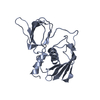
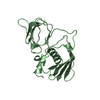
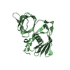
- Structure visualization
| Structure viewer | Molecule: MolmilJmol/JSmol |
|---|
- Downloads & links
-Download
| PDBx/mmCIF format | 1aw7.cif.gz | 168.6 KB | Display | PDBx/mmCIF format |
|---|---|---|---|---|
| PDB format | pdb1aw7.ent.gz | 134.2 KB | Display | PDB format |
| PDBx/mmJSON format | 1aw7.json.gz | Tree view | PDBx/mmJSON format | |
| Others |  Other downloads Other downloads |
-Validation report
| Arichive directory | https://data.pdbj.org/pub/pdb/validation_reports/aw/1aw7ftp://data.pdbj.org/pub/pdb/validation_reports/aw/1aw7 | HTTPS FTP |
|---|
-Related structure data
| Related structure data |  1ts2C  1ts3C  1ts4C  1ts5C  1tss S: Starting model for refinement C: citing same article ( |
|---|---|
| Similar structure data |


-Links
 PDBj
PDBj
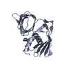
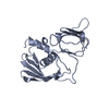
- Assembly
| Deposited unit | 
| ||||||||||||||||
|---|---|---|---|---|---|---|---|---|---|---|---|---|---|---|---|---|---|
| 1 | 
| ||||||||||||||||
| 2 | 
| ||||||||||||||||
| 3 | 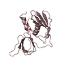
| ||||||||||||||||
| 4 | 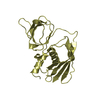
| ||||||||||||||||
| Unit cell |
| ||||||||||||||||
| Noncrystallographic symmetry (NCS) | NCS oper:
|
-Components
| #1: Protein | Mass: 22046.719 Da / Num. of mol.: 4 Mutation: CHAIN A, Q136A, CHAIN B, Q336A, CHAIN C, Q536A, CHAIN D, Q736A Source method: isolated from a genetically manipulated source Source: (gene. exp.) Staphylococcus aureus (bacteria) / References: UniProt: P06886#2: Water | ChemComp-HOH / |  Mass: 18.015 Da / Num. of mol.: 291 / Source method: isolated from a natural source / Formula: H2O Mass: 18.015 Da / Num. of mol.: 291 / Source method: isolated from a natural source / Formula: H2O |
|---|
-Experimental details
-Experiment
| Experiment | Method: X-RAY DIFFRACTION / Number of used crystals: 1 |
|---|
- Sample preparation
| Crystal | Density Matthews: 2.3 Å3/Da / Density % sol: 46.6 % | |||||||||||||||||||||||||
|---|---|---|---|---|---|---|---|---|---|---|---|---|---|---|---|---|---|---|---|---|---|---|---|---|---|---|
| Crystal grow | pH: 7.5 / Details: pH 7.5 | |||||||||||||||||||||||||
| Crystal grow | *PLUS Method: vapor diffusion | |||||||||||||||||||||||||
| Components of the solutions | *PLUS
|
-Data collection
| Diffraction | Mean temperature: 293 K |
|---|---|
| Diffraction source | Source: ROTATING ANODE / Type: RIGAKU RUH2R / Wavelength: 1.5418 |
| Detector | Type: SIEMENS / Detector: AREA DETECTOR / Date: Nov 1, 1996 |
| Radiation | Monochromator: SI(111) / Monochromatic (M) / Laue (L): M / Scattering type: x-ray |
| Radiation wavelength | Wavelength: 1.5418 Å / Relative weight: 1 |
| Reflection | Resolution: 2.3→20 Å / Num. obs: 38992 / % possible obs: 79 % / Redundancy: 3.1 % / Rsym value: 0.056 / Net I/σ(I): 12.7 |
| Reflection shell | Resolution: 1.95→2.01 Å / Redundancy: 1.3 % / Mean I/σ(I) obs: 1 / Rsym value: 0.24 / % possible all: 37 |
| Reflection | *PLUS Highest resolution: 1.95 Å / Num. obs: 50028 / Num. measured all: 157168 / Rmerge(I) obs: 0.056 |
- Processing
| Software |
| ||||||||||||||||||||||||||||||||||||||||||||||||||||||||||||||||||||||||||||||||
|---|---|---|---|---|---|---|---|---|---|---|---|---|---|---|---|---|---|---|---|---|---|---|---|---|---|---|---|---|---|---|---|---|---|---|---|---|---|---|---|---|---|---|---|---|---|---|---|---|---|---|---|---|---|---|---|---|---|---|---|---|---|---|---|---|---|---|---|---|---|---|---|---|---|---|---|---|---|---|---|---|---|
| Refinement | Method to determine structure: MOLECULAR REPLACEMENT Starting model: PDB ENTRY 1TSS 1tss Resolution: 1.95→20 Å / Data cutoff high absF: 100000000 / Data cutoff low absF: 0 / σ(F): 0 /
| ||||||||||||||||||||||||||||||||||||||||||||||||||||||||||||||||||||||||||||||||
| Displacement parameters | Biso mean: 25.27 Å2 | ||||||||||||||||||||||||||||||||||||||||||||||||||||||||||||||||||||||||||||||||
| Refine analyze | Luzzati coordinate error obs: 0.25 Å | ||||||||||||||||||||||||||||||||||||||||||||||||||||||||||||||||||||||||||||||||
| Refinement step | Cycle: LAST / Resolution: 1.95→20 Å
| ||||||||||||||||||||||||||||||||||||||||||||||||||||||||||||||||||||||||||||||||
| Refine LS restraints |
| ||||||||||||||||||||||||||||||||||||||||||||||||||||||||||||||||||||||||||||||||
| LS refinement shell | Resolution: 1.95→1.98 Å / Rfactor Rwork: 0.319 / Total num. of bins used: 20 |
